Summary information and primary citation
- PDB-id
- 4qyz; SNAP-derived features in text and JSON formats;
DNAproDB
- Class
- immune system-DNA-RNA
- Method
- X-ray (3.03 Å)
- Summary
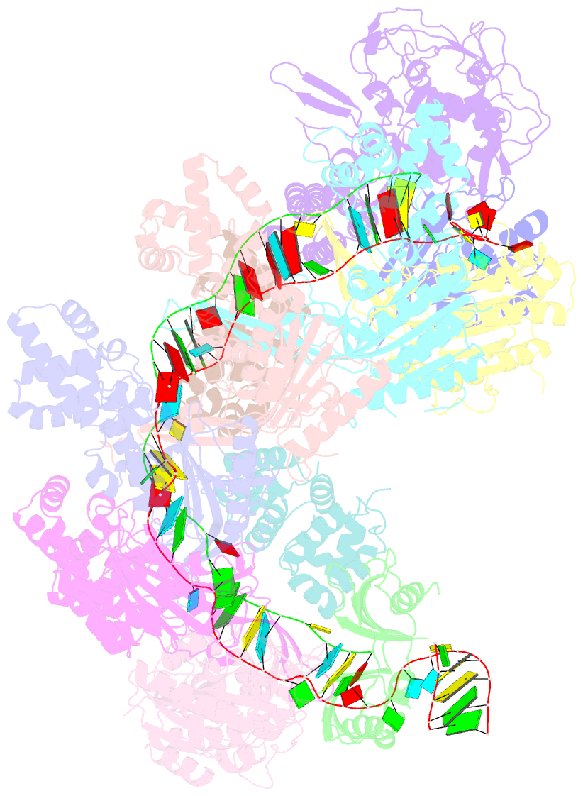
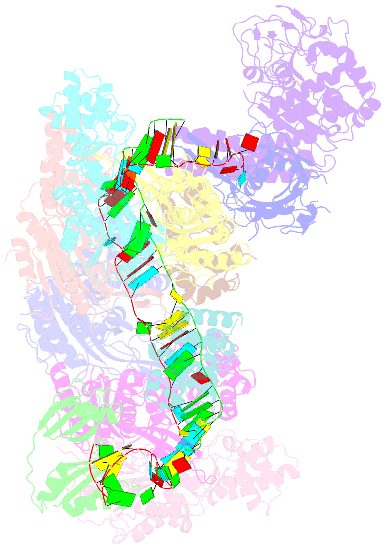
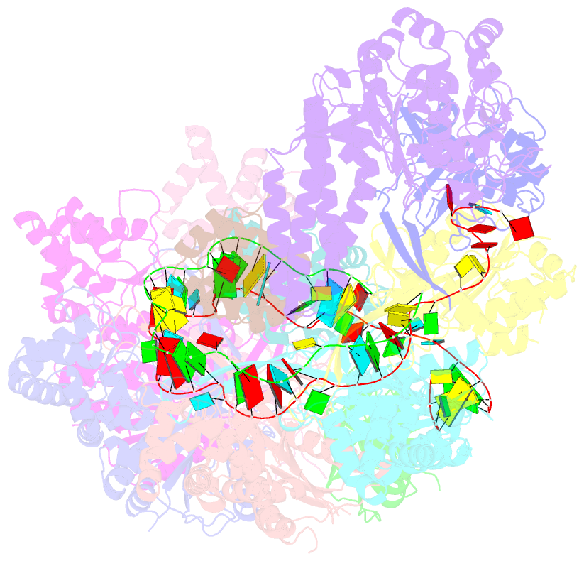
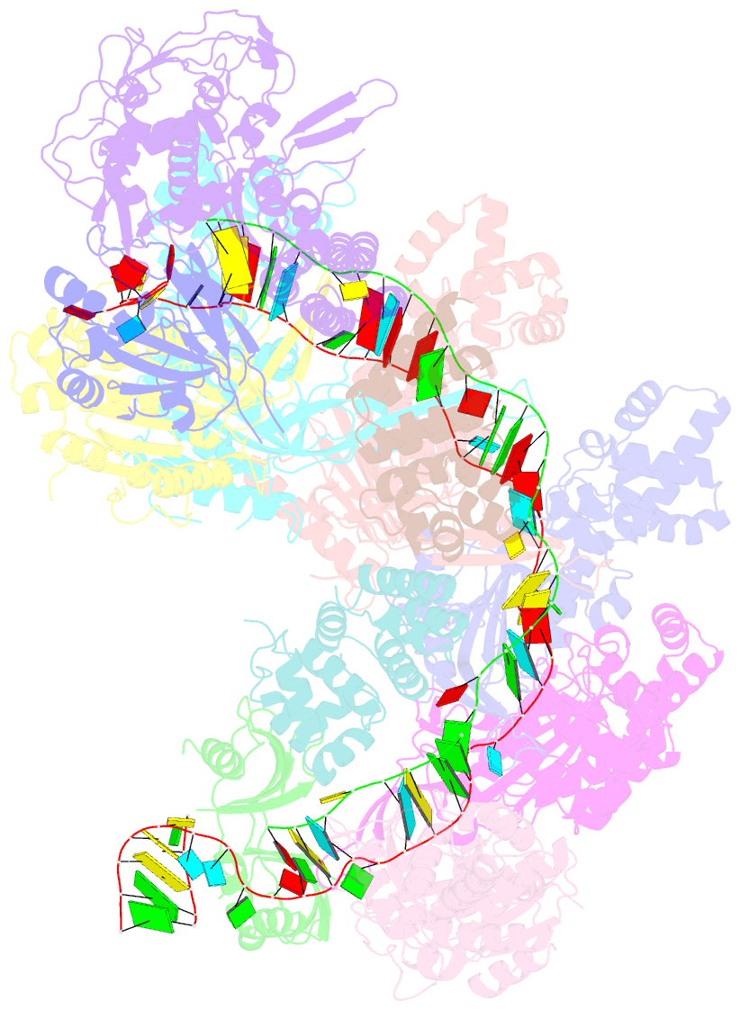
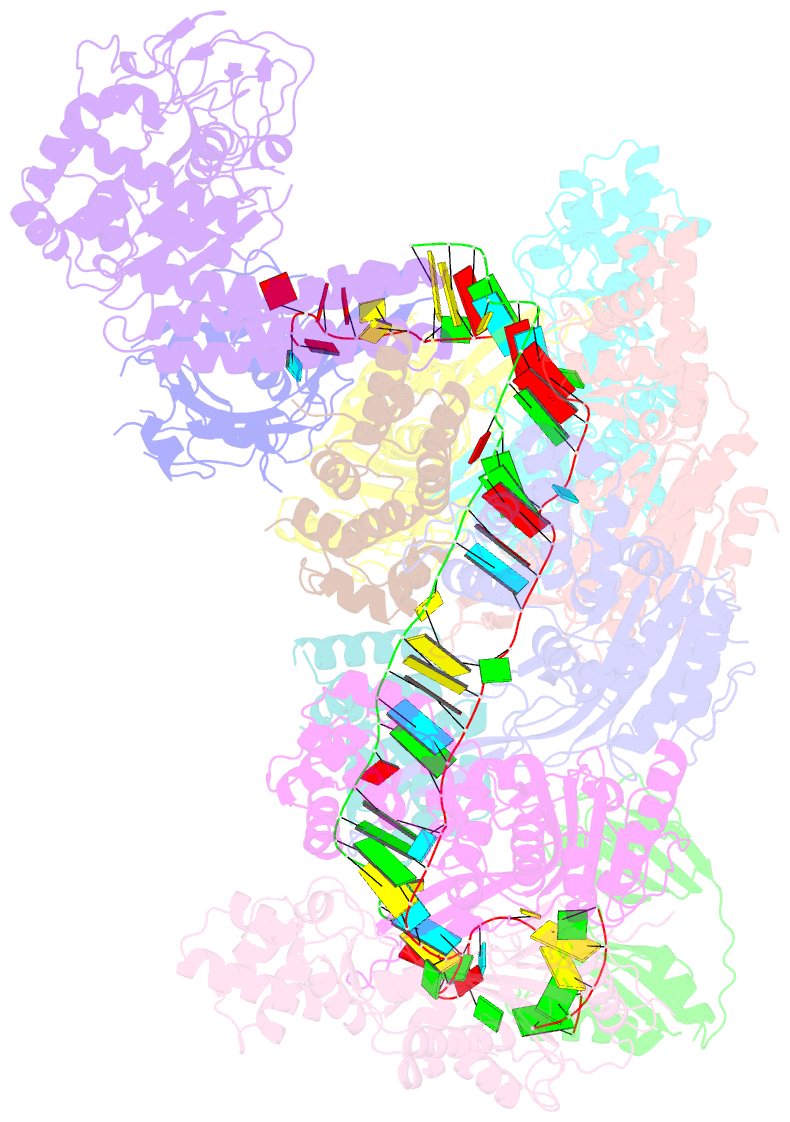
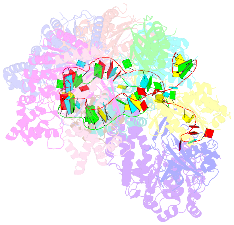
- Crystal structure of a crispr RNA-guided surveillance complex, cascade, bound to a ssDNA target
- Reference
- Mulepati S, Heroux A, Bailey S (2014): "Structural biology. Crystal structure of a CRISPR RNA-guided surveillance complex bound to a ssDNA target." Science, 345, 1479-1484. doi: 10.1126/science.1256996.
- Abstract
- In prokaryotes, RNA derived from type I and type III CRISPR loci direct large ribonucleoprotein complexes to destroy invading bacteriophage and plasmids. In Escherichia coli, this 405-kilodalton complex is called Cascade. We report the crystal structure of Cascade bound to a single-stranded DNA (ssDNA) target at a resolution of 3.03 angstroms. The structure reveals that the CRISPR RNA and target strands do not form a double helix but instead adopt an underwound ribbon-like structure. This noncanonical structure is facilitated by rotation of every sixth nucleotide out of the RNA-DNA hybrid and is stabilized by the highly interlocked organization of protein subunits. These studies provide insight into both the assembly and the activity of this complex and suggest a mechanism to enforce fidelity of target binding.