Summary information and primary citation
- PDB-id
- 4rq5; SNAP-derived features in text and JSON formats;
DNAproDB
- Class
- transferase-DNA
- Method
- X-ray (2.32 Å)
- Summary
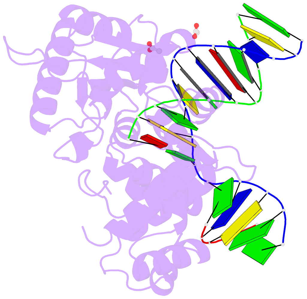
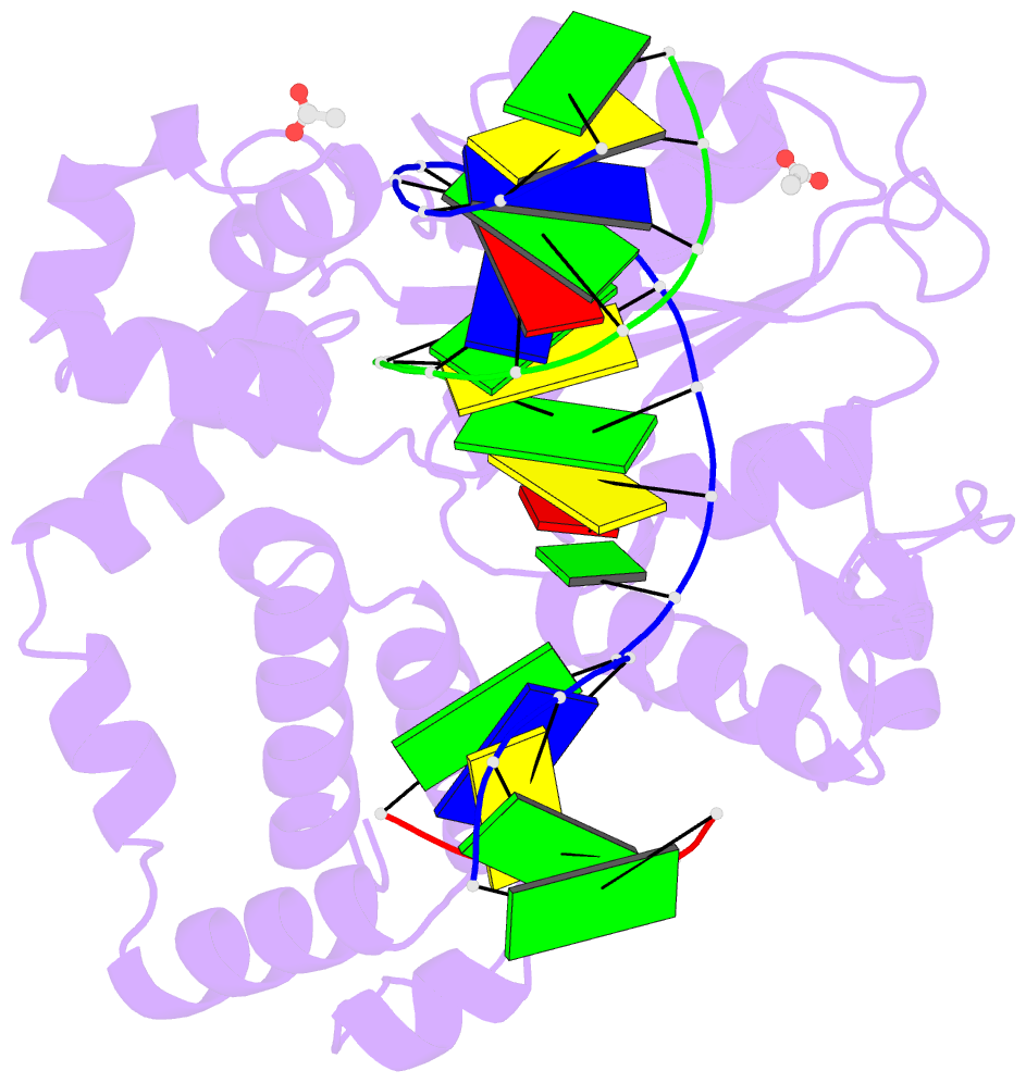
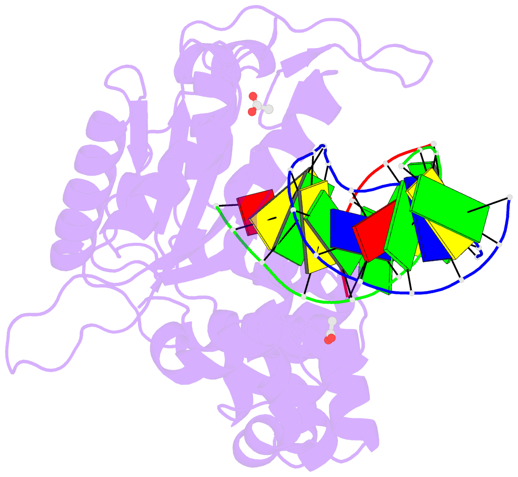
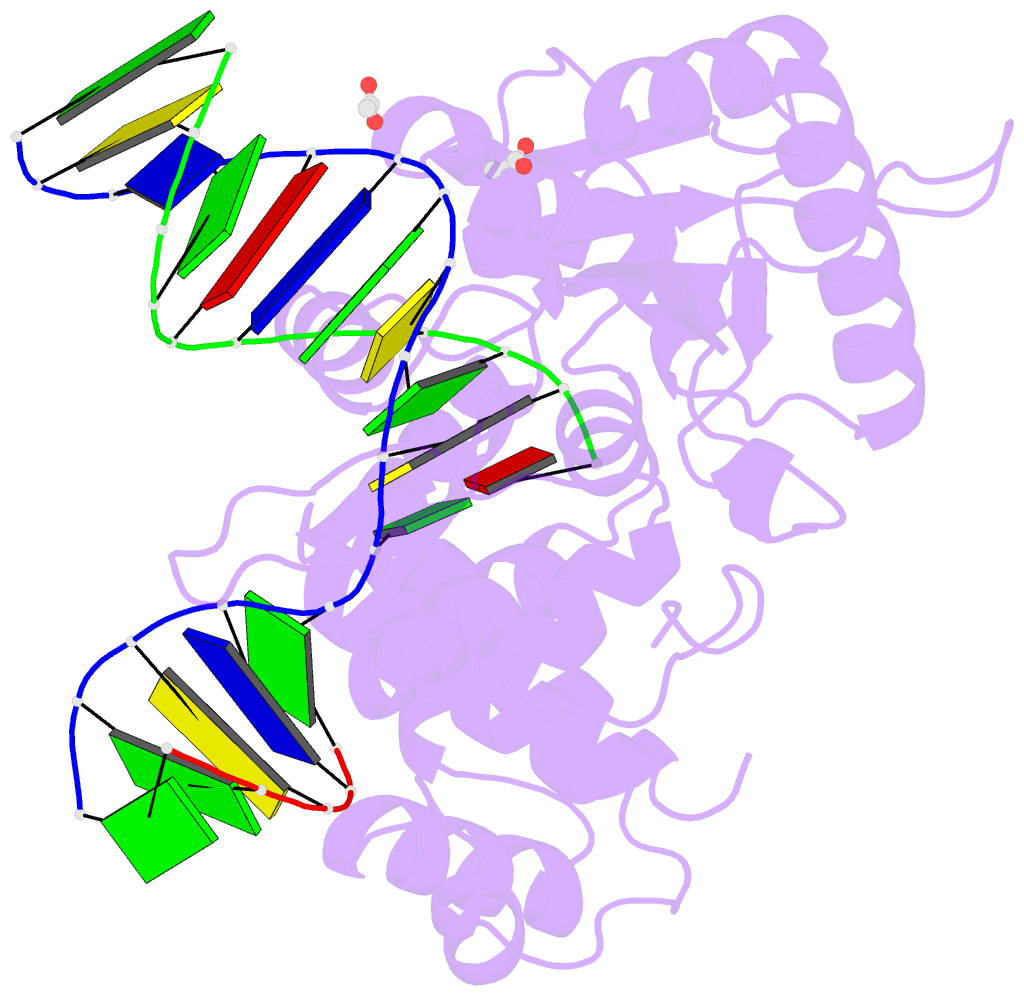
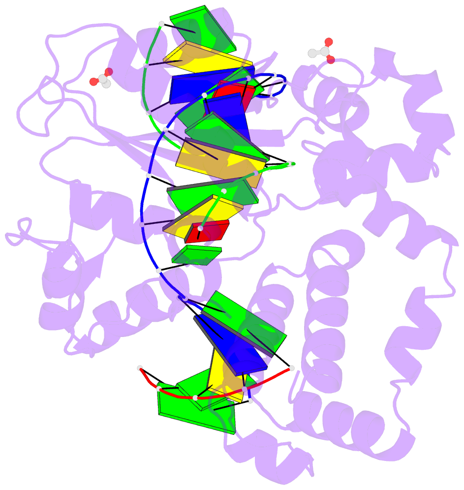
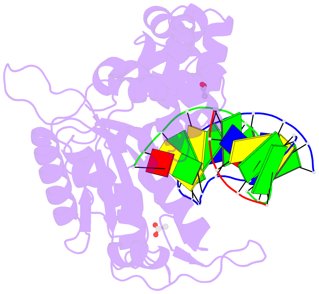
- Human DNA polymerase beta with gapped DNA containing an 8-oxo-7,8-dihydro-guanine (8-oxog)and datp soaked with mgcl2 for 60 s
- Reference
- Vyas R, Reed AJ, Tokarsky EJ, Suo Z (2015): "Viewing Human DNA Polymerase beta Faithfully and Unfaithfully Bypass an Oxidative Lesion by Time-Dependent Crystallography." J.Am.Chem.Soc., 137, 5225-5230. doi: 10.1021/jacs.5b02109.
- Abstract
- One common oxidative DNA lesion, 8-oxo-7,8-dihydro-2'-deoxyguanine (8-oxoG), is highly mutagenic in vivo due to its anti-conformation forming a Watson-Crick base pair with correct deoxycytidine 5'-triphosphate (dCTP) and its syn-conformation forming a Hoogsteen base pair with incorrect deoxyadenosine 5'-triphosphate (dATP). Here, we utilized time-resolved X-ray crystallography to follow 8-oxoG bypass by human DNA polymerase β (hPolβ). In the 12 solved structures, both Watson-Crick (anti-8-oxoG:anti-dCTP) and Hoogsteen (syn-8-oxoG:anti-dATP) base pairing were clearly visible and were maintained throughout the chemical reaction. Additionally, a third Mg(2+) appeared during the process of phosphodiester bond formation and was located between the reacting α- and β-phosphates of the dNTP, suggesting its role in stabilizing reaction intermediates. After phosphodiester bond formation, hPolβ reopened its conformation, pyrophosphate was released, and the newly incorporated primer 3'-terminal nucleotide stacked, rather than base paired, with 8-oxoG. These structures provide the first real-time pictures, to our knowledge, of how a polymerase correctly and incorrectly bypasses a DNA lesion.