Summary information and primary citation
- PDB-id
- 4uqm; SNAP-derived features in text and JSON formats;
DNAproDB
- Class
- hydrolase-DNA
- Method
- X-ray (1.35 Å)
- Summary
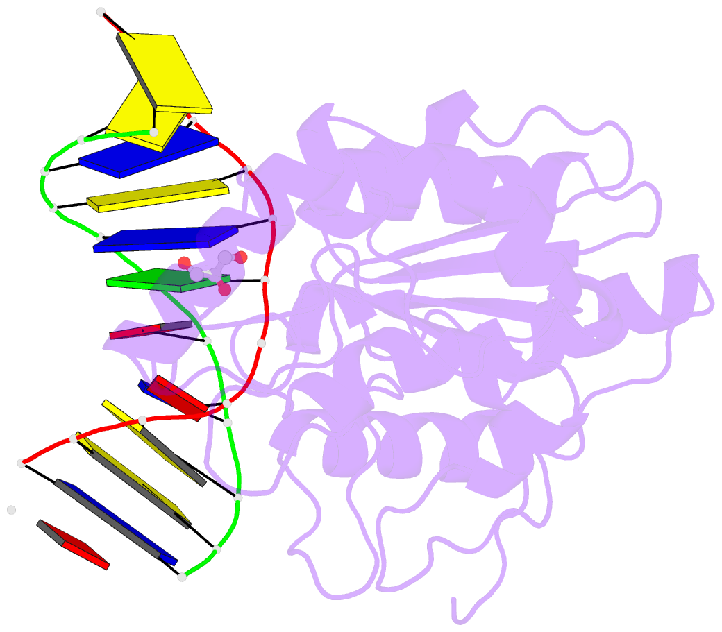
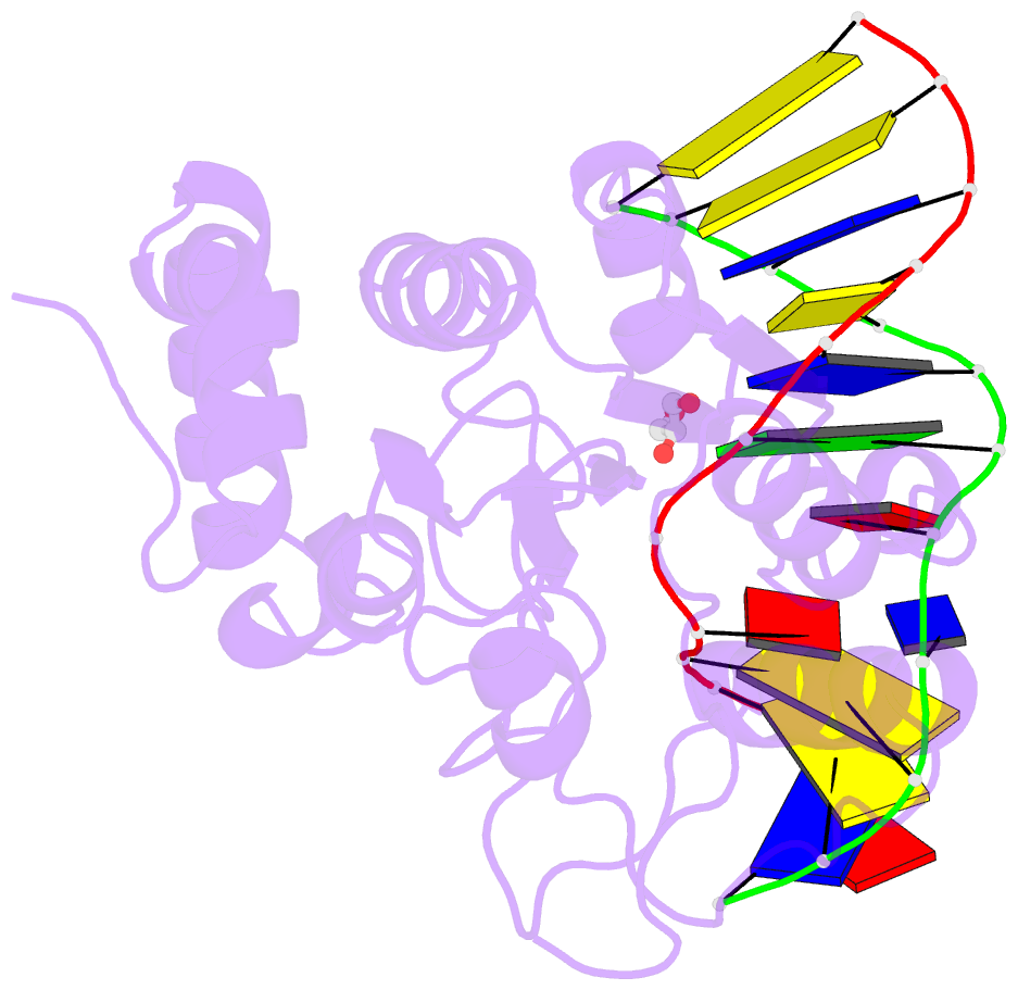
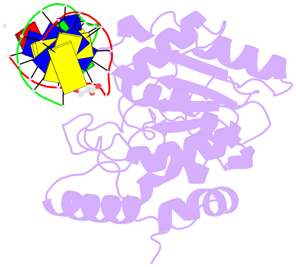
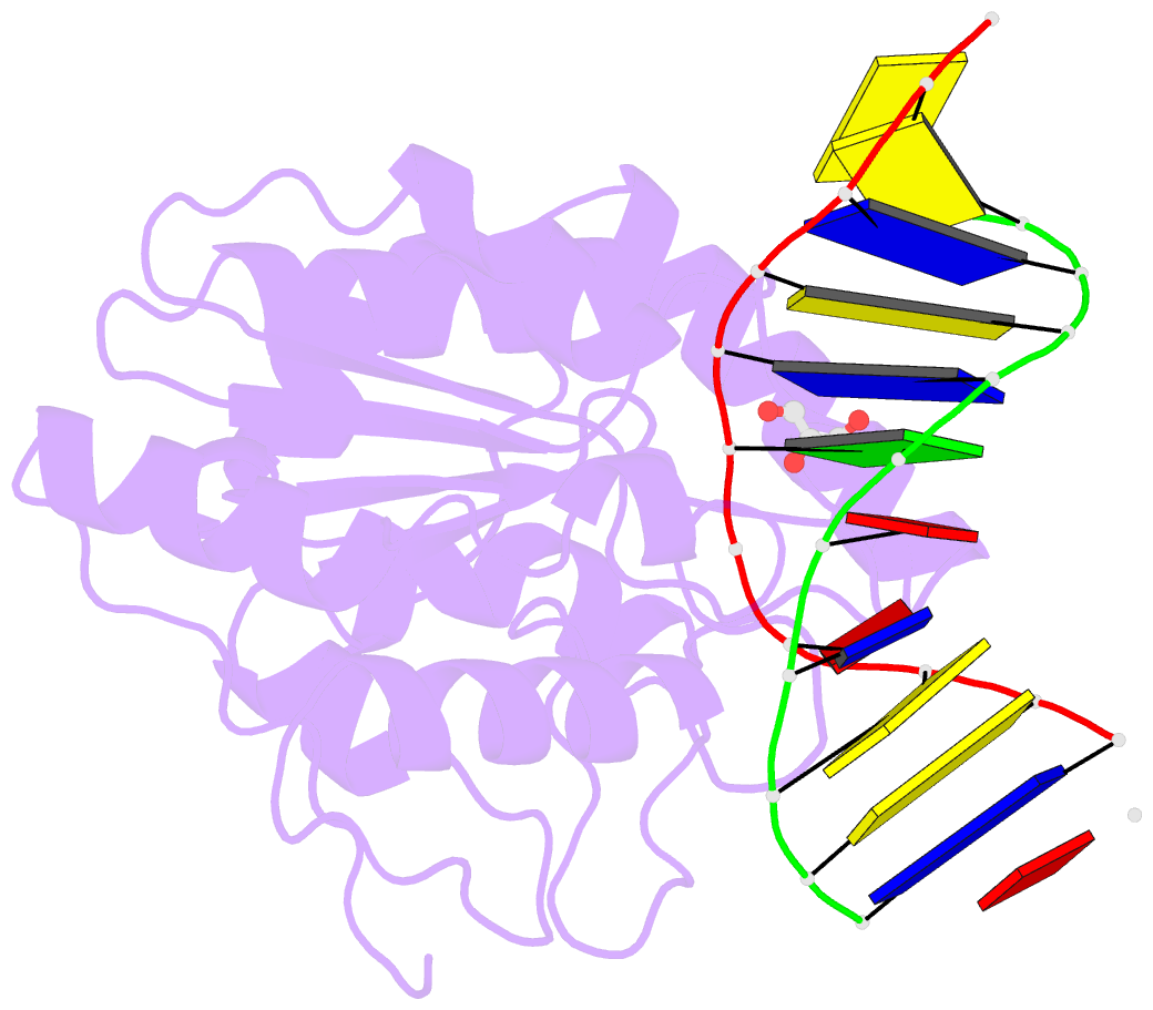
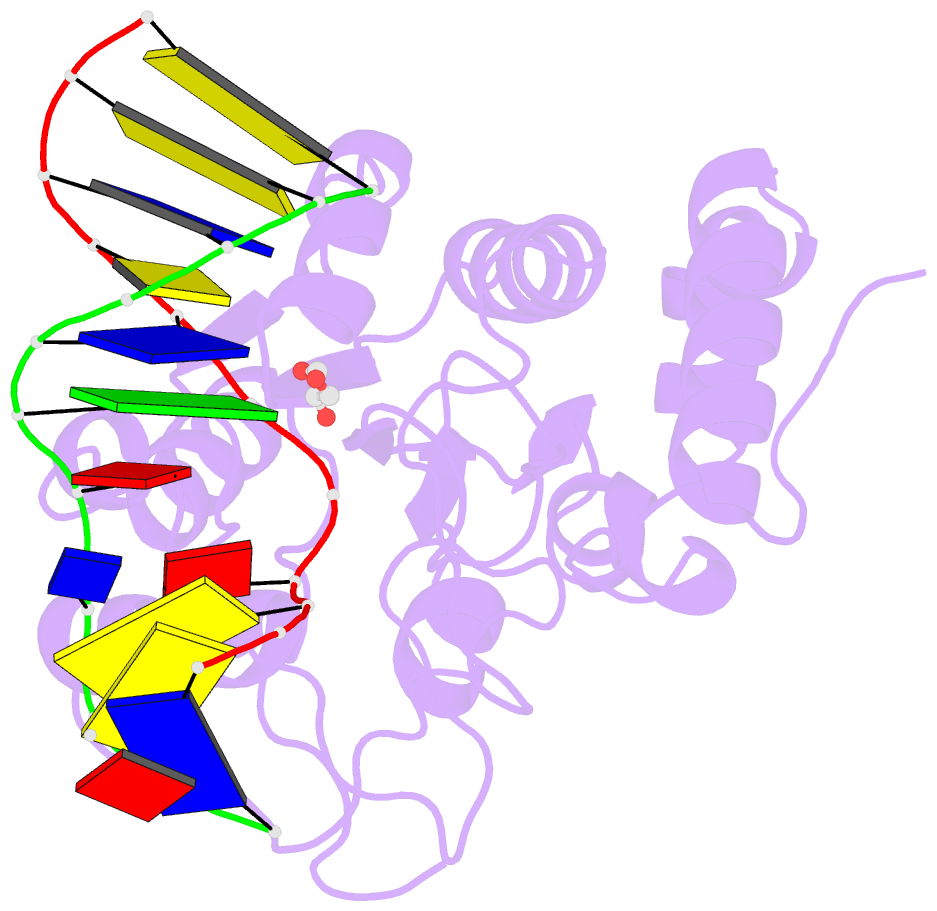
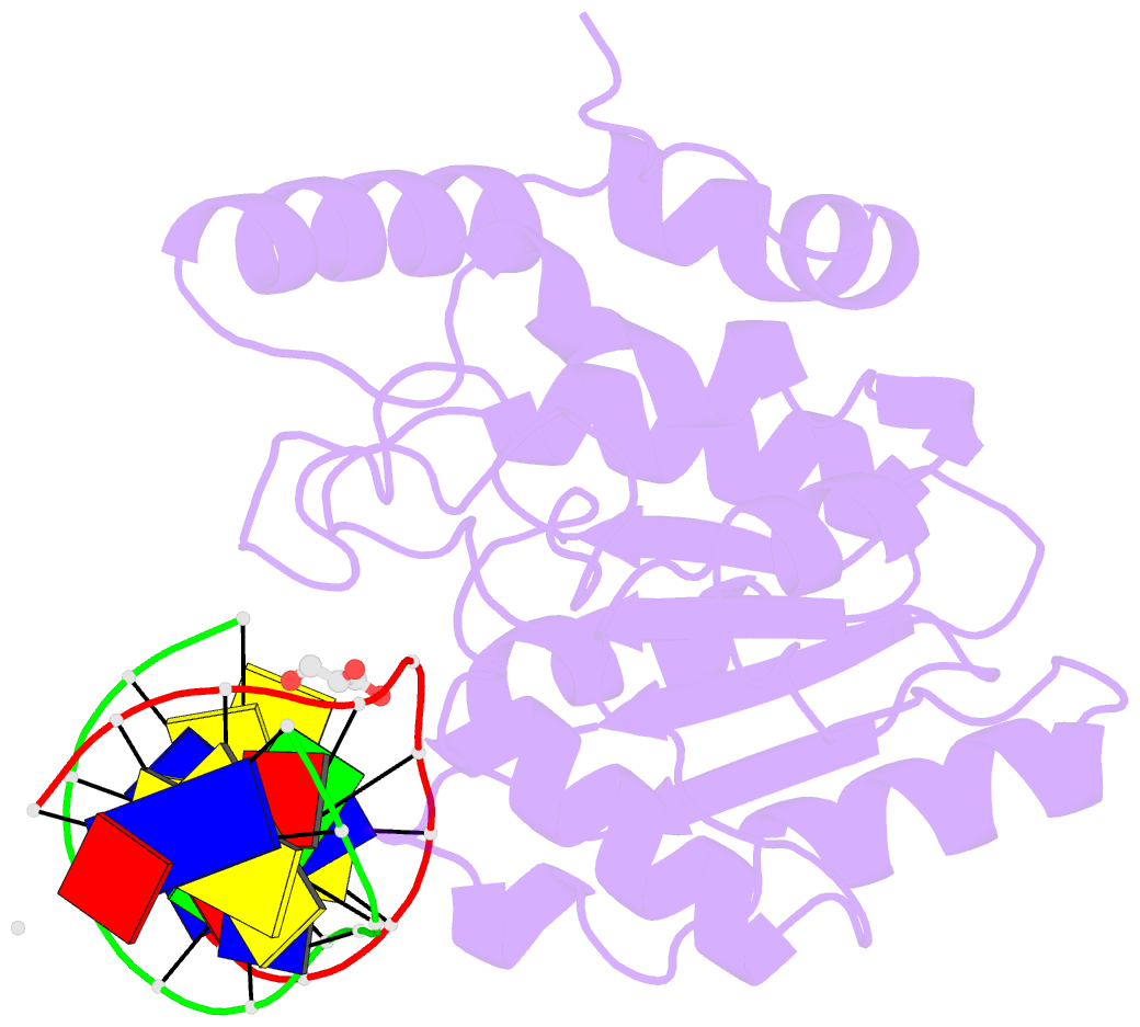
- Crystal structure determination of uracil-DNA n-glycosylase (ung) from deinococcus radiodurans in complex with DNA - new insights into the role of the leucine-loop for damage recognition and repair
- Reference
- Pedersen HL, Johnson KA, McVey CE, Leiros I, Moe E (2015): "Structure determination of uracil-DNA N-glycosylase from Deinococcus radiodurans in complex with DNA." Acta Crystallogr. D Biol. Crystallogr., 71, 2137-2149. doi: 10.1107/S1399004715014157.
- Abstract
- Uracil-DNA N-glycosylase (UNG) is a DNA-repair enzyme in the base-excision repair (BER) pathway which removes uracil from DNA. Here, the crystal structure of UNG from the extremophilic bacterium Deinococcus radiodurans (DrUNG) in complex with DNA is reported at a resolution of 1.35 Å. Prior to the crystallization experiments, the affinity between DrUNG and different DNA oligonucleotides was tested by electrophoretic mobility shift assays (EMSAs). As a result of this analysis, two 16 nt double-stranded DNAs were chosen for the co-crystallization experiments, one of which (16 nt AU) resulted in well diffracting crystals. The DNA in the co-crystal structure contained an abasic site (substrate product) flipped into the active site of the enzyme, with no uracil in the active-site pocket. Despite the high resolution, it was not possible to fit all of the terminal nucleotides of the DNA complex into electron density owing to disorder caused by a lack of stabilizing interactions. However, the DNA which was in contact with the enzyme, close to the active site, was well ordered and allowed detailed analysis of the enzyme-DNA interaction. The complex revealed that the interaction between DrUNG and DNA is similar to that in the previously determined crystal structure of human UNG (hUNG) in complex with DNA [Slupphaug et al. (1996). Nature (London), 384, 87-92]. Substitutions in a (here defined) variable part of the leucine loop result in a shorter loop (eight residues instead of nine) in DrUNG compared with hUNG; regardless of this, it seems to fulfil its role and generate a stabilizing force with the minor groove upon flipping out of the damaged base into the active site. The structure also provides a rationale for the previously observed high catalytic efficiency of DrUNG caused by high substrate affinity by demonstrating an increased number of long-range electrostatic interactions between the enzyme and the DNA. Interestingly, specific interactions between residues in the N-terminus of a symmetry-related molecule and the complementary DNA strand facing away from the active site were also observed which seem to stabilize the enzyme-DNA complex. However, the significance of this observation remains to be investigated. The results provide new insights into the current knowledge about DNA damage recognition and repair by uracil-DNA glycosylases.