Summary information and primary citation
- PDB-id
- 4uuv; SNAP-derived features in text and JSON formats;
DNAproDB
- Class
- transcription
- Method
- X-ray (2.8 Å)
- Summary
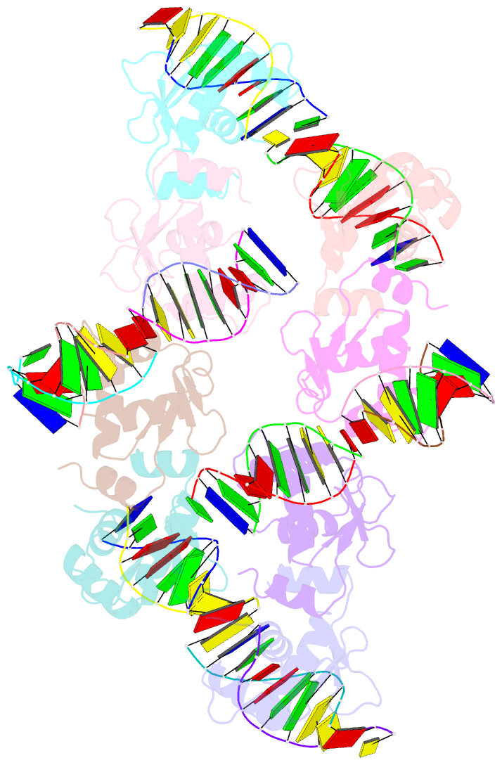
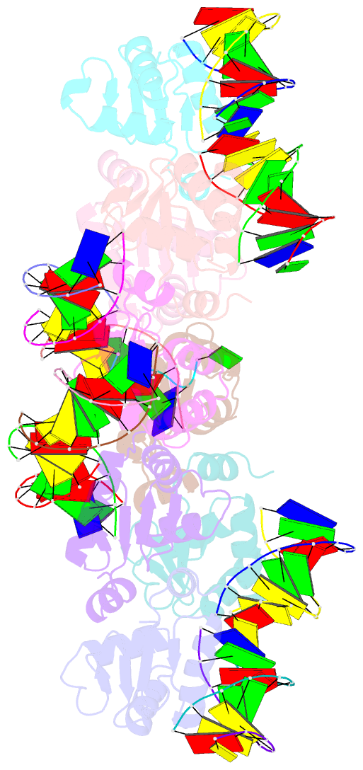
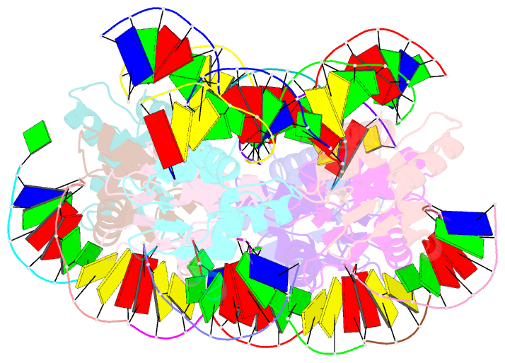
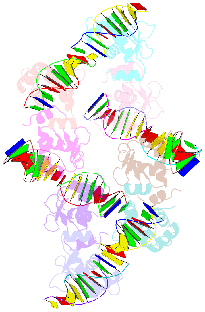
- Structure of the DNA binding ets domain of human etv4 in complex with DNA
- Reference
- Cooper CDO, Newman JA, Aitkenhead H, Allerston CK, Gileadi O (2015): "Structures of the Ets Domains of Transcription Factors Etv1, Etv4, Etv5 and Fev: Determinants of DNA Binding and Redox Regulation by Disulfide Bond Formation." J.Biol.Chem., 290, 13692. doi: 10.1074/JBC.M115.646737.
- Abstract
- Ets transcription factors, which share the conserved Ets DNA-binding domain, number nearly 30 members in humans and are particularly involved in developmental processes. Their deregulation following changes in expression, transcriptional activity, or by chromosomal translocation plays a critical role in carcinogenesis. Ets DNA binding, selectivity, and regulation have been extensively studied; however, questions still arise regarding binding specificity outside the core GGA recognition sequence and the mode of action of Ets post-translational modifications. Here, we report the crystal structures of Etv1, Etv4, Etv5, and Fev, alone and in complex with DNA. We identify previously unrecognized features of the protein-DNA interface. Interactions with the DNA backbone account for most of the binding affinity. We describe a highly coordinated network of water molecules acting in base selection upstream of the GGAA core and the structural features that may account for discrimination against methylated cytidine residues. Unexpectedly, all proteins crystallized as disulfide-linked dimers, exhibiting a novel interface (distant to the DNA recognition helix). Homodimers of Etv1, Etv4, and Etv5 could be reduced to monomers, leading to a 40-200-fold increase in DNA binding affinity. Hence, we present the first indication of a redox-dependent regulatory mechanism that may control the activity of this subset of oncogenic Ets transcription factors.