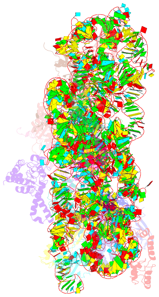
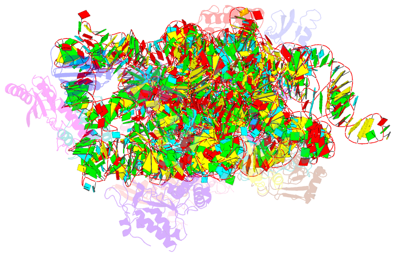
Summary information and primary citation
- PDB-id
- 4yhh; SNAP-derived features in text and JSON formats;
DNAproDB
- Class
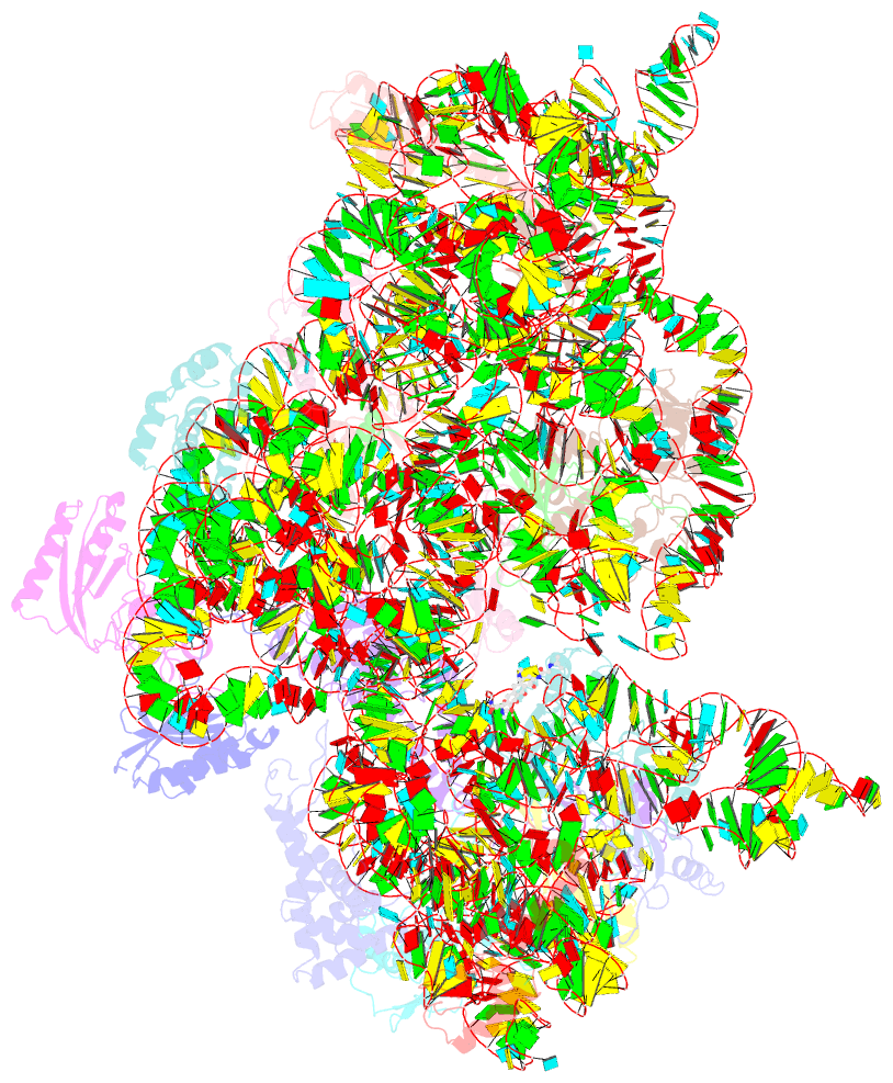
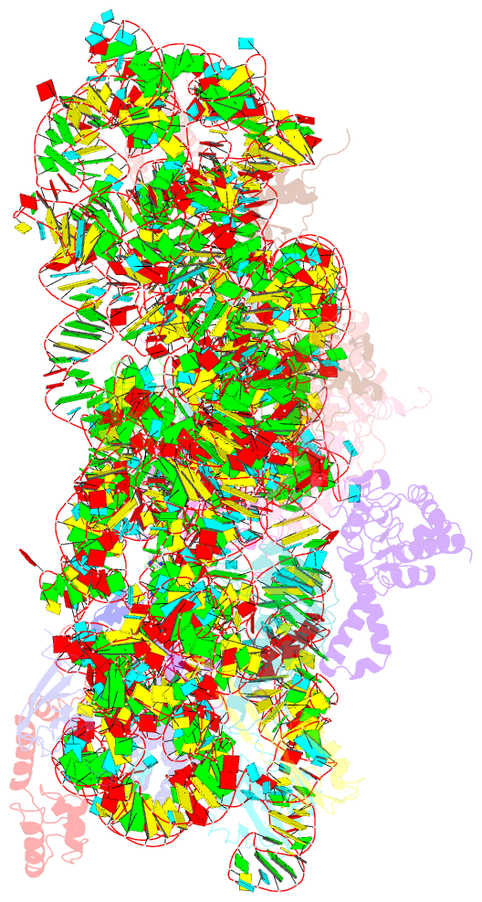
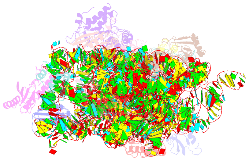
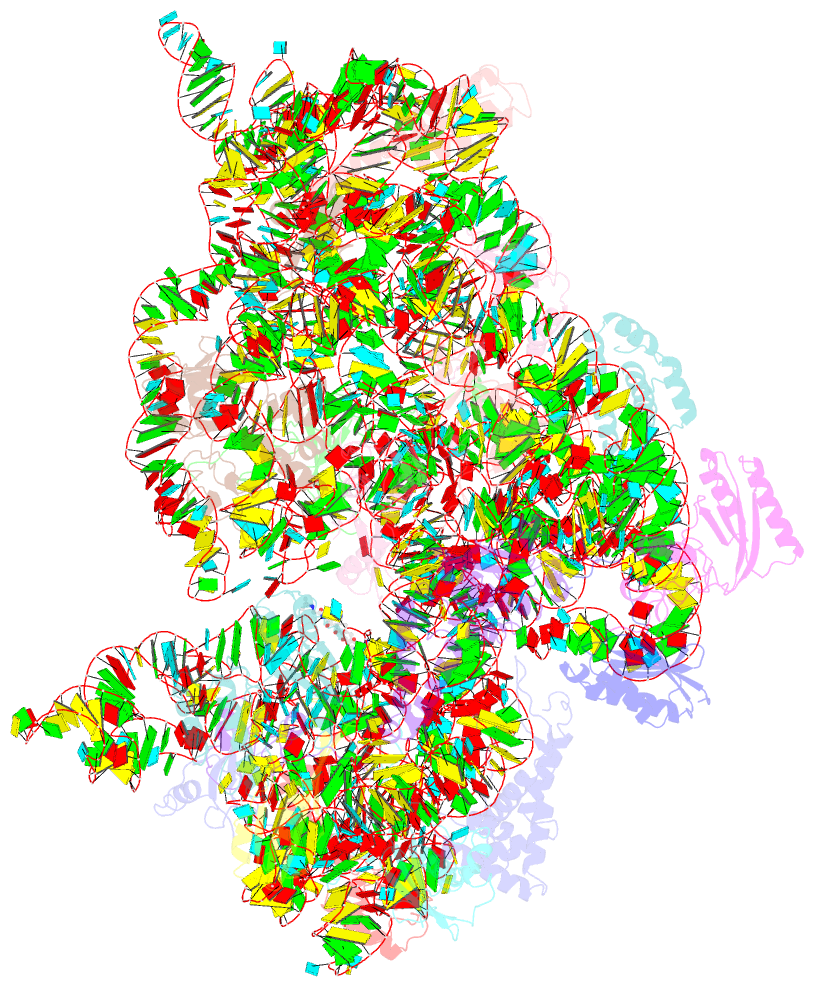
- ribosome
- Method
- X-ray (3.417 Å)
- Summary
- Crystal structure of the 30s ribosomal subunit from thermus thermophilus in complex with tigecycline
- Reference
- Schedlbauer A, Kaminishi T, Ochoa-Lizarralde B, Dhimole N, Zhou S, Lopez-Alonso JP, Connell SR, Fucini P (2015): "Structural characterization of an alternative mode of tigecycline binding to the bacterial ribosome." Antimicrob.Agents Chemother., 59, 2849-2854. doi: 10.1128/AAC.04895-14.
- Abstract
- Although both tetracycline and tigecycline inhibit protein synthesis by sterically hindering the binding of tRNA to the ribosomal A site, tigecycline shows increased efficacy in both in vitro and in vivo activity assays and escapes the most common resistance mechanisms associated with the tetracycline class of antibiotics. These differences in activities are attributed to the tert-butyl-glycylamido side chain found in tigecycline. Our structural analysis by X-ray crystallography shows that tigecycline binds the bacterial 30S ribosomal subunit with its tail in an extended conformation and makes extensive interactions with the 16S rRNA nucleotide C1054. These interactions restrict the mobility of C1054 and contribute to the antimicrobial activity of tigecycline, including its resistance to the ribosomal protection proteins.