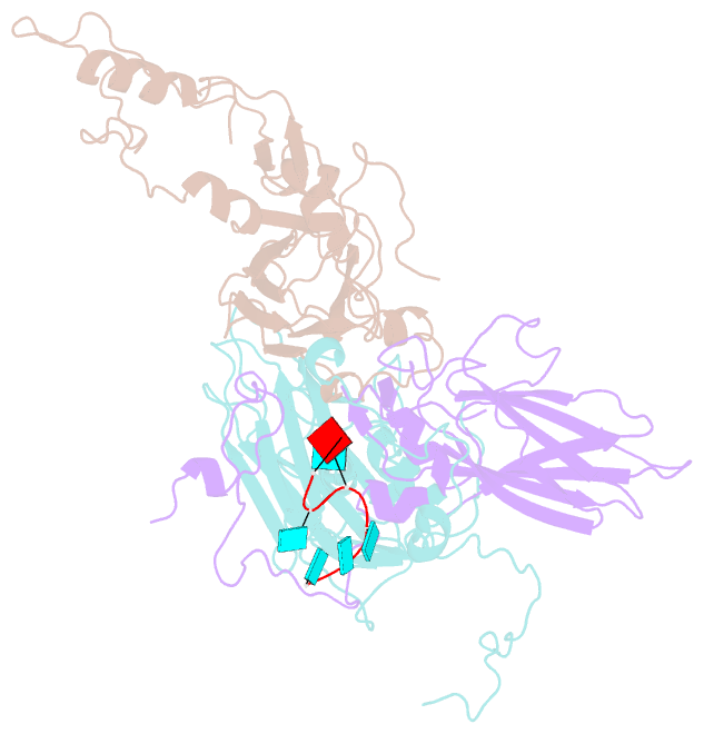
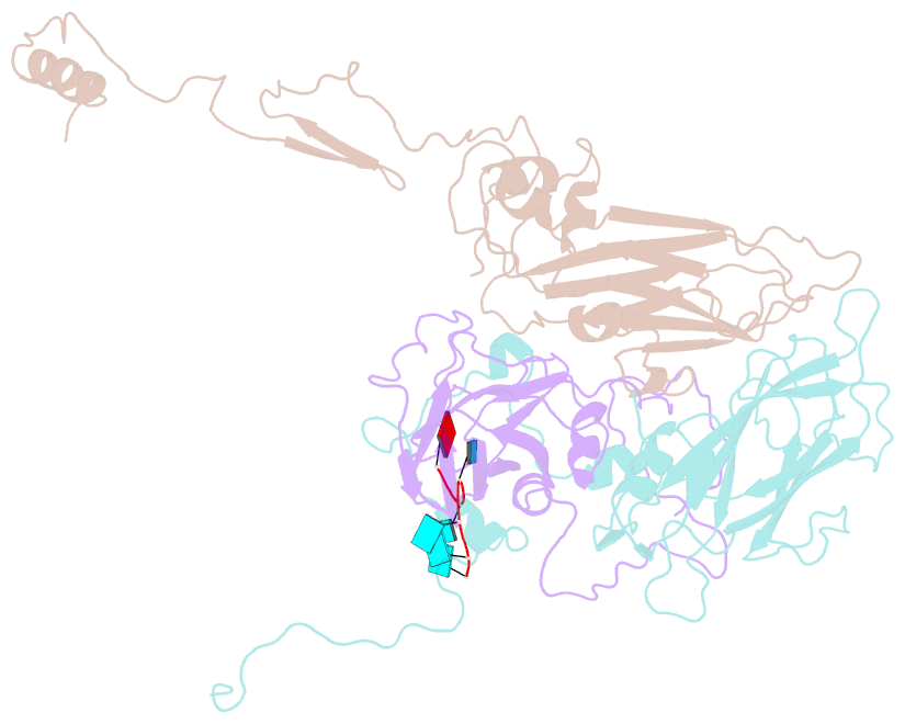
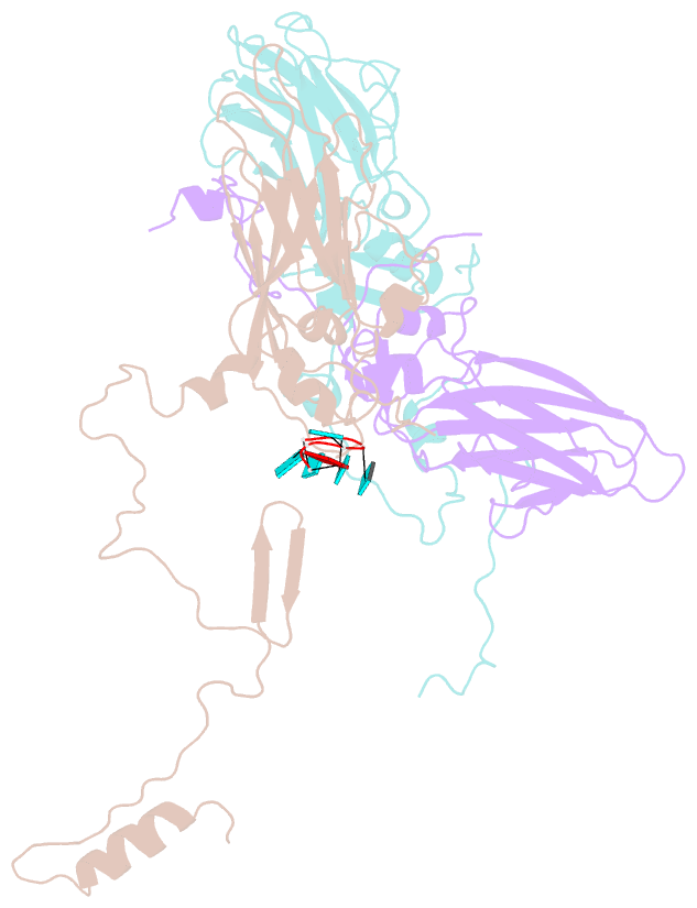
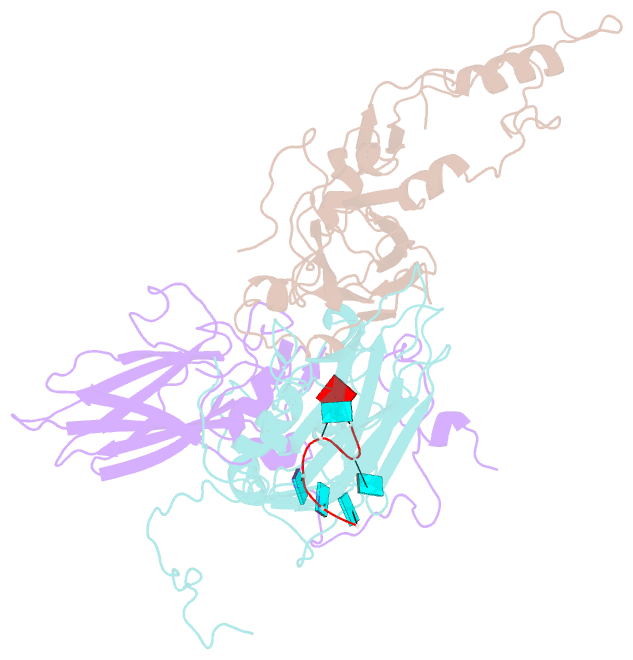
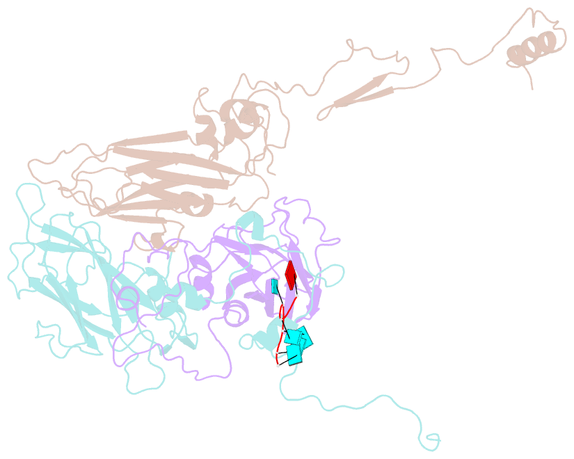
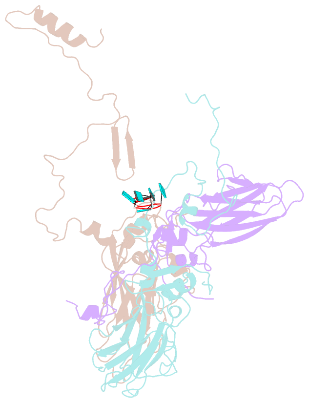
Summary information and primary citation

PDB-id
4z92; SNAP-derived features in text and JSON formats; DNAproDB
Class
virus
Method
X-ray (3.1 Å)
Summary
Crystal structure of parechovirus-1 virion
Reference
Kalynych S, Palkova L, Plevka P (2015): "The Structure of Human Parechovirus 1 Reveals an Association of the RNA Genome with the Capsid." J.Virol., 90, 1377-1386. doi: 10.1128/JVI.02346-15.
Abstract
Parechoviruses are human pathogens that cause diseases ranging from gastrointestinal disorders to encephalitis. Unlike most picornaviruses, parechovirus capsids are composed of only three subunits: VP0, VP1, and VP3. Here we present the structure of a human parechovirus-1 (HPeV-1) virion determined to a resolution of 3.1 Å. We find that interactions among pentamers in the HPeV-1 capsid are mediated by the N-termini of VP0s, which correspond to the capsid protein VP4 and the N-terminal part of the capsid protein VP2 of other picornaviruses. In order to facilitate delivery of the virus genome into the cytoplasm, the N-termini of VP0s have to be released from contacts between pentamers and exposed at the particle surface, resulting in capsid disruption. A hydrophobic pocket, which can be targeted by capsid-binding anti-viral compounds in many other picornaviruses, is not present in HPeV-1. However, we found that interactions between the HPeV-1 ssRNA genome and subunits VP1 and VP3 in the virion impose a partial icosahedral ordering on the genome. The residues involved in the RNA binding are conserved among all parechoviruses, suggesting putative role of the genome in virion stability or assembly. Therefore, putative small molecules that could disrupt HPeV RNA-capsid protein interactions could be developed into antiviral inhibitors.

Cartoon-block schematics in six views (download the tarball)

PyMOL session file Download PDB file View in 3Dmol.js