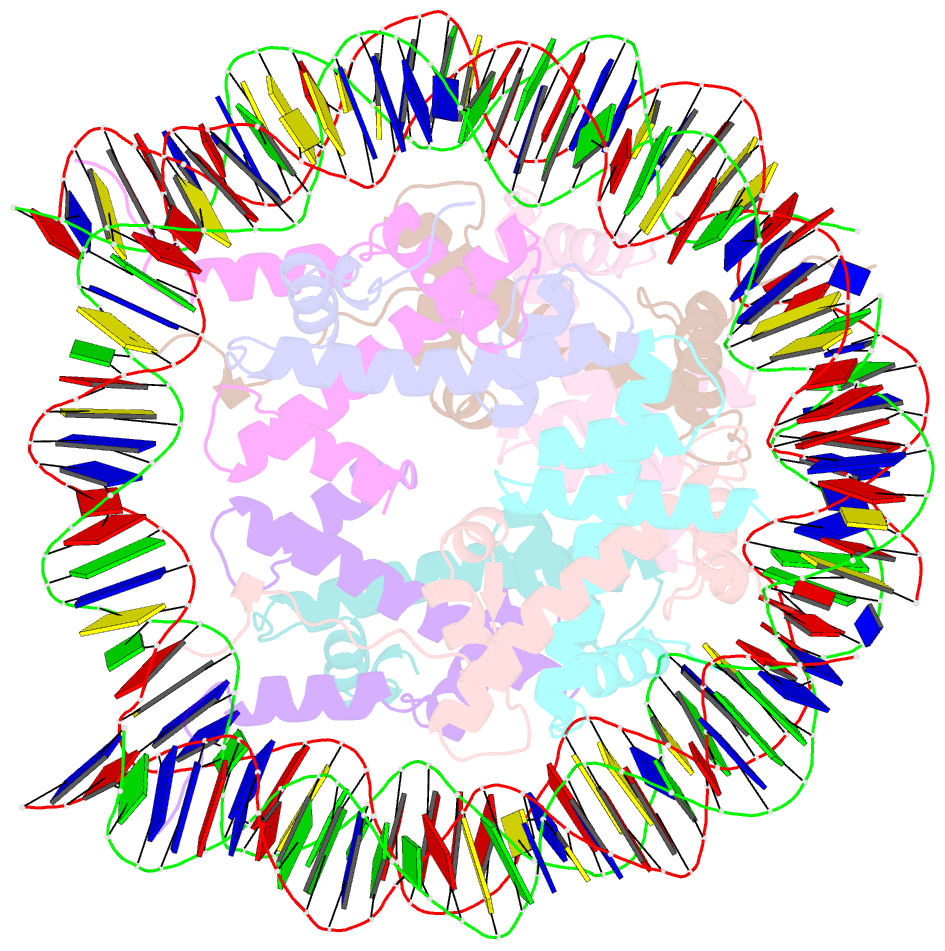
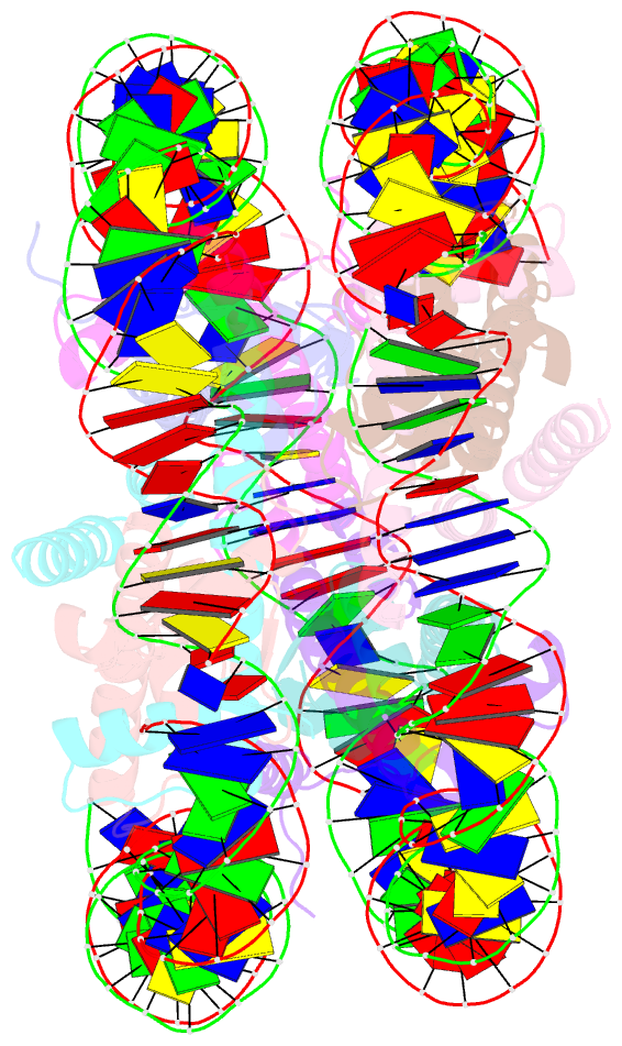
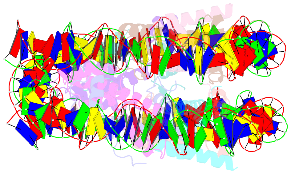
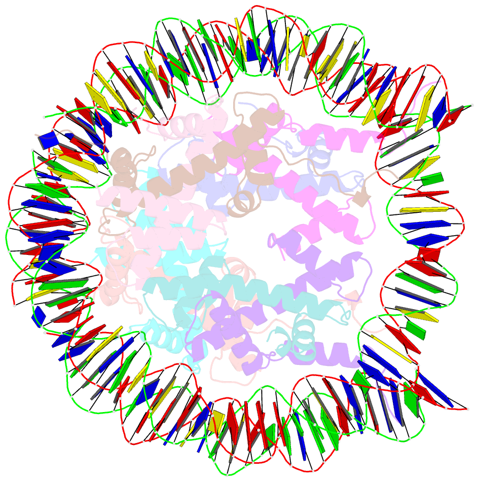
Summary information and primary citation
- PDB-id
- 5b24; SNAP-derived features in text and JSON formats;
DNAproDB
- Class
- structural protein-DNA
- Method
- X-ray (3.6 Å)
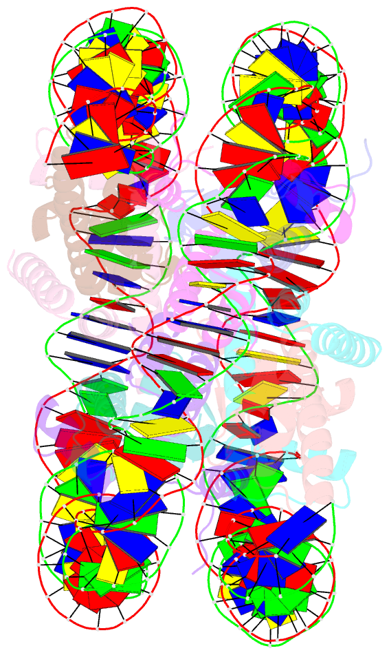
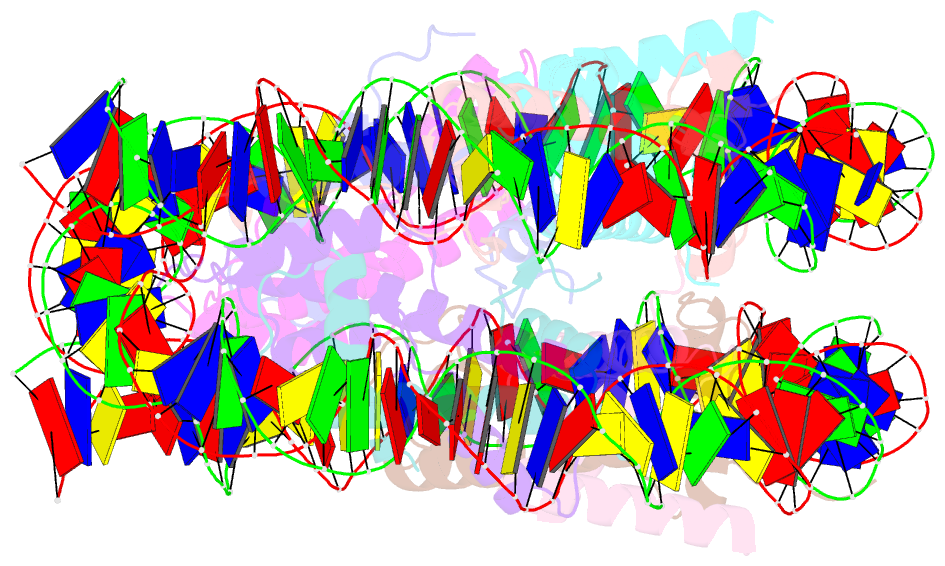
- Summary
- The crystal structure of the nucleosome containing cyclobutane pyrimidine dimer
- Reference
- Horikoshi N, Tachiwana H, Kagawa W, Osakabe A, Matsumoto S, Iwai S, Sugasawa K, Kurumizaka H (2016): "Crystal structure of the nucleosome containing ultraviolet light-induced cyclobutane pyrimidine dimer." Biochem.Biophys.Res.Commun., 471, 117-122. doi: 10.1016/j.bbrc.2016.01.170.
- Abstract
- The cyclobutane pyrimidine dimer (CPD) is induced in genomic DNA by ultraviolet (UV) light. In mammals, this photolesion is primarily induced within nucleosomal DNA, and repaired exclusively by the nucleotide excision repair (NER) pathway. However, the mechanism by which the CPD is accommodated within the nucleosome has remained unknown. We now report the crystal structure of a nucleosome containing CPDs. In the nucleosome, the CPD induces only limited local backbone distortion, and the affected bases are accommodated within the duplex. Interestingly, one of the affected thymine bases is located within 3.0 Å from the undamaged complementary adenine base, suggesting the formation of complementary hydrogen bonds in the nucleosome. We also found that UV-DDB, which binds the CPD at the initial stage of the NER pathway, also efficiently binds to the nucleosomal CPD. These results provide important structural and biochemical information for understanding how the CPD is accommodated and recognized in chromatin.