Summary information and primary citation
- PDB-id
- 5f6c; SNAP-derived features in text and JSON formats;
DNAproDB
- Class
- hydrolase
- Method
- X-ray (3.002 Å)
- Summary
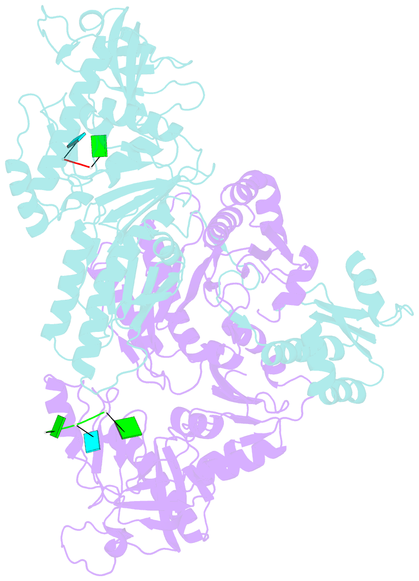
- The structure of e. coli rnase e catalytically inactive mutant with RNA bound
- Reference
- Bandyra KJ, Wandzik JM, Luisi BF (2018): "Substrate Recognition and Autoinhibition in the Central Ribonuclease RNase E." Mol. Cell, 72, 275-285.e4. doi: 10.1016/j.molcel.2018.08.039.
- Abstract
- The endoribonuclease RNase E is a principal factor in RNA turnover and processing that helps to exercise fine control of gene expression in bacteria. While its catalytic activity can be strongly influenced by the chemical identity of the 5' end of RNA substrates, the enzyme can also cleave numerous substrates irrespective of the chemistry of their 5' ends through a mechanism that has remained largely unexplained. We report structural and functional data illuminating details of both operational modes. Our crystal structure of RNase E in complex with the sRNA RprA reveals a duplex recognition site that saddles an inter-protomer surface to help present substrates for cleavage. Our data also reveal an autoinhibitory pocket that modulates the overall activity of the ribonuclease. Taking these findings together, we propose how RNase E uses versatile modes of RNA recognition to achieve optimal activity and specificity.


