Summary information and primary citation
- PDB-id
- 5fmz; SNAP-derived features in text and JSON formats;
DNAproDB
- Class
- transcription
- Method
- X-ray (3.4 Å)
- Summary
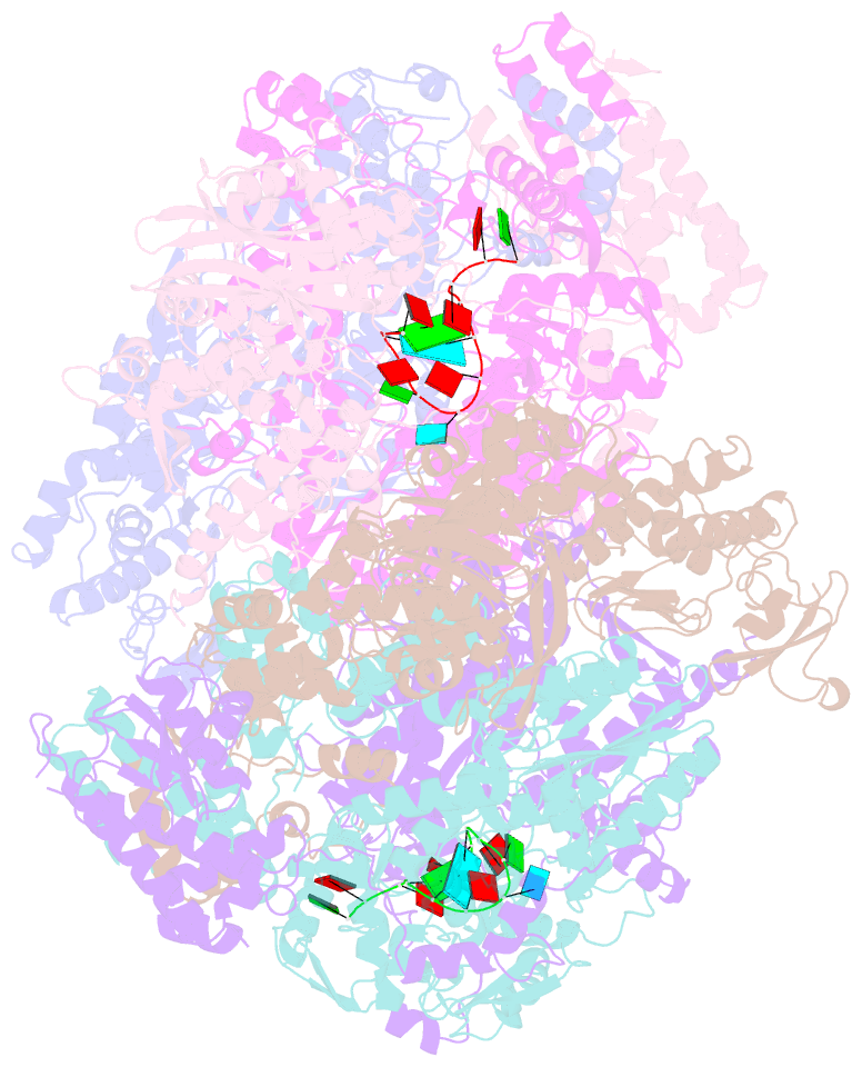
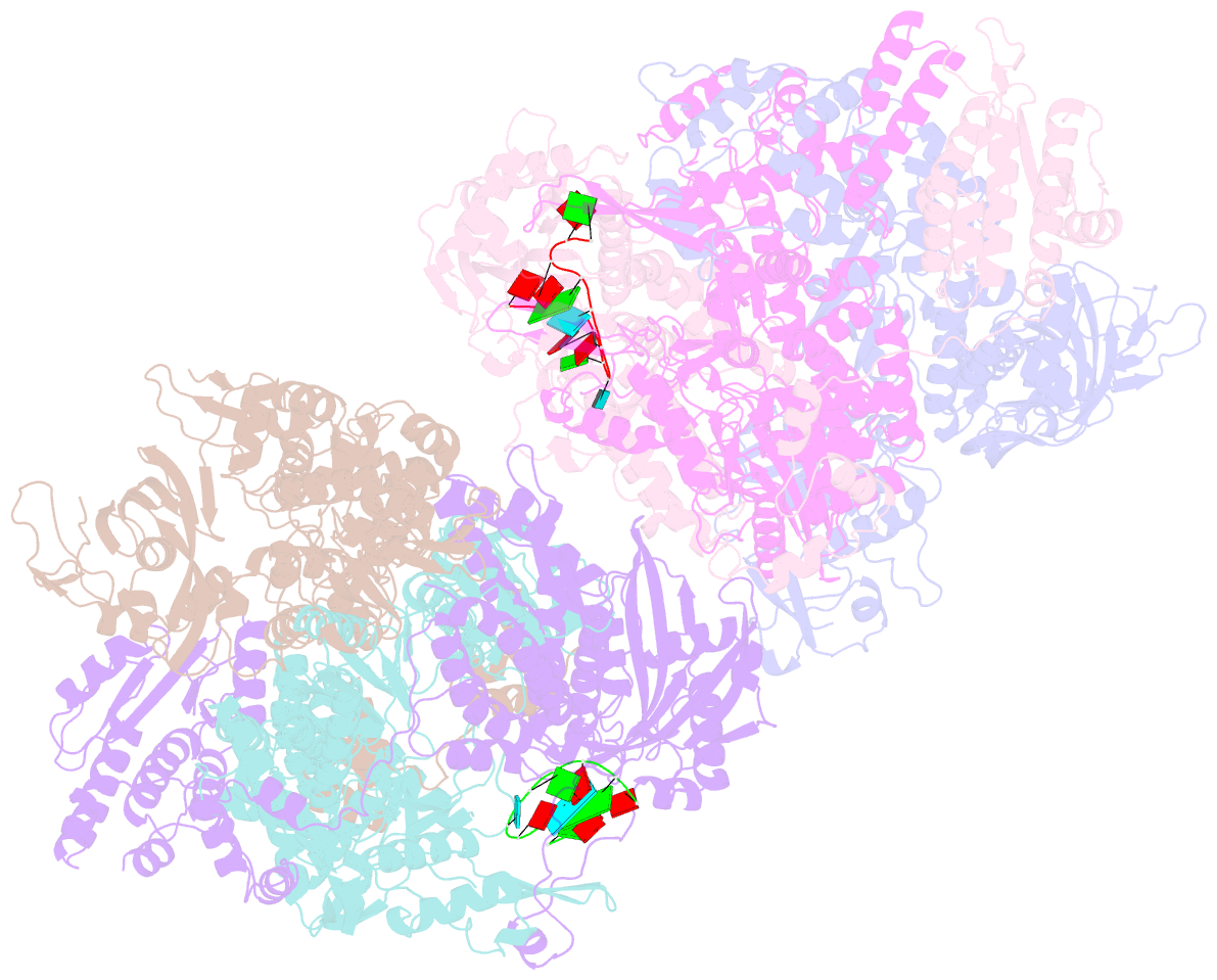
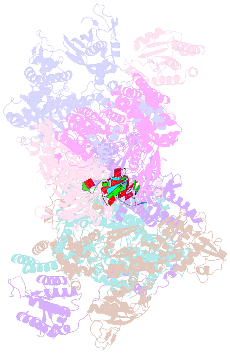
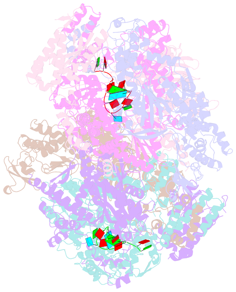
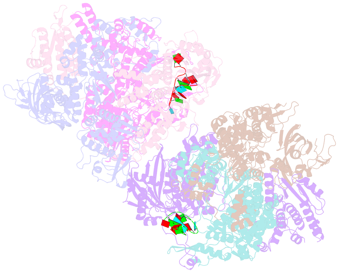
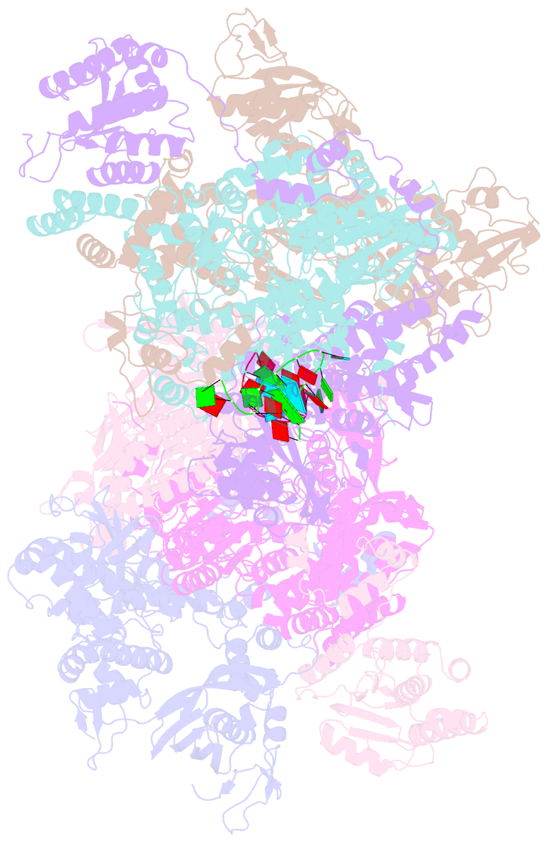
- Crystal structure of influenza b polymerase with bound 5' vrna
- Reference
- Thierry E, Guilligay D, Kosinski J, Bock T, Gaudon S, Round A, Pflug A, Hengrung N, El Omari K, Baudin F, Hart DJ, Beck M, Cusack S (2016): "Influenza Polymerase Can Adopt an Alternative Configuration Involving a Radical Repacking of Pb2 Domains." Mol.Cell, 61, 125. doi: 10.1016/J.MOLCEL.2015.11.016.
- Abstract
- Influenza virus polymerase transcribes or replicates the segmented RNA genome (vRNA) into respectively viral mRNA or full-length copies and initiates RNA synthesis by binding the conserved 3' and 5' vRNA ends (the promoter). In recent structures of promoter-bound polymerase, the cap-binding and endonuclease domains are configured for cap snatching, which generates capped transcription primers. Here, we present a FluB polymerase structure with a bound complementary cRNA 5' end that exhibits a major rearrangement of the subdomains within the C-terminal two-thirds of PB2 (PB2-C). Notably, the PB2 nuclear localization signal (NLS)-containing domain translocates ∼90 Å to bind to the endonuclease domain. FluA PB2-C alone and RNA-free FluC polymerase are similarly arranged. Biophysical and cap-dependent endonuclease assays show that in solution the polymerase explores different conformational distributions depending on which RNA is bound. The inherent flexibility of the polymerase allows it to adopt alternative conformations that are likely important during polymerase maturation into active progeny RNPs.