Summary information and primary citation
- PDB-id
- 5n8l; SNAP-derived features in text and JSON formats;
DNAproDB
- Class
- RNA binding protein
- Method
- NMR
- Summary
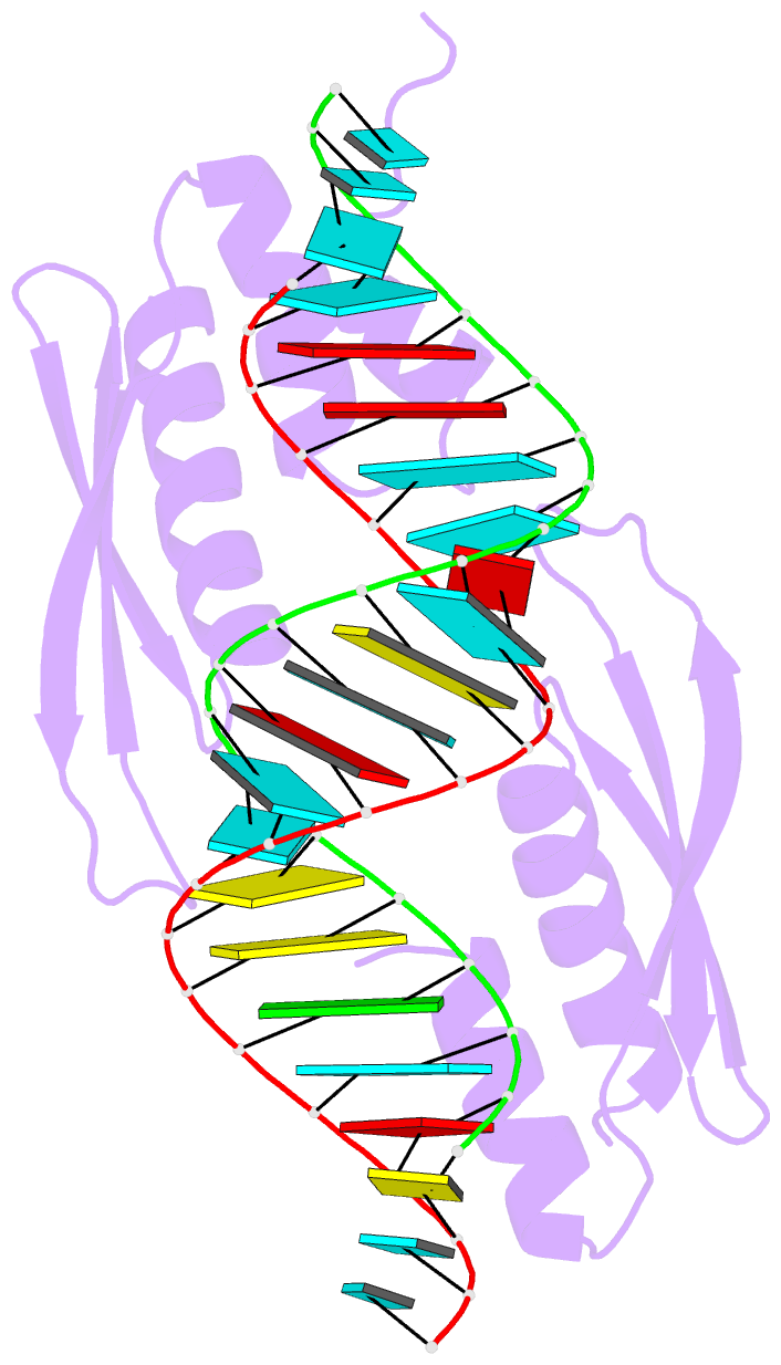
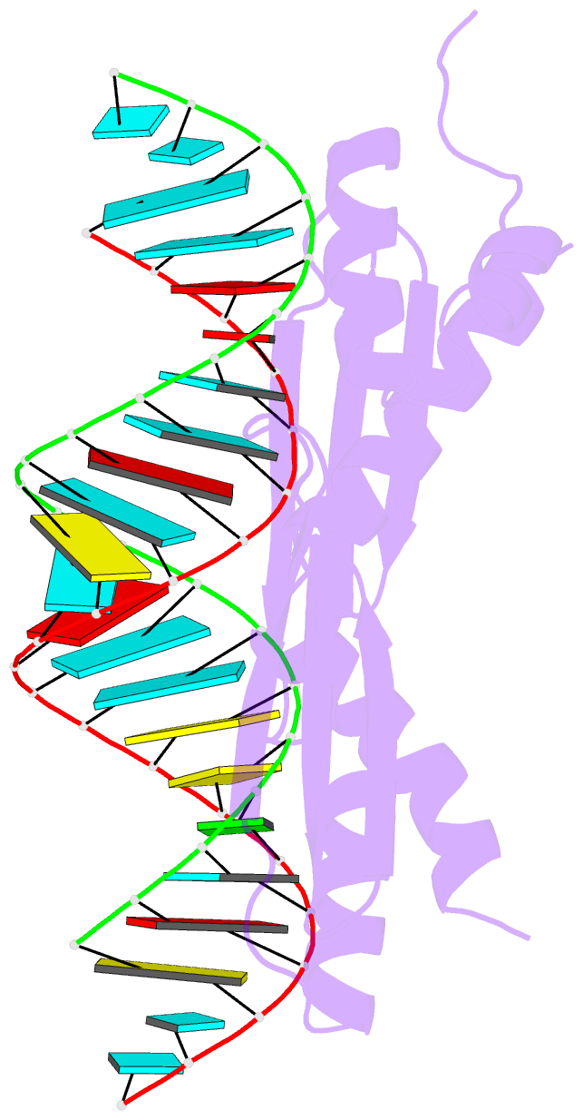
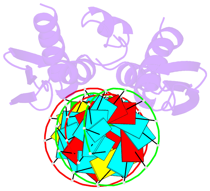
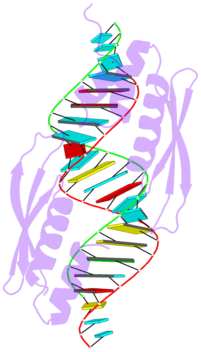
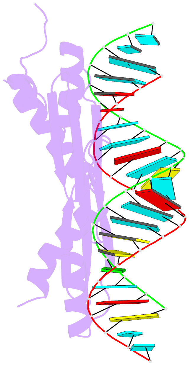
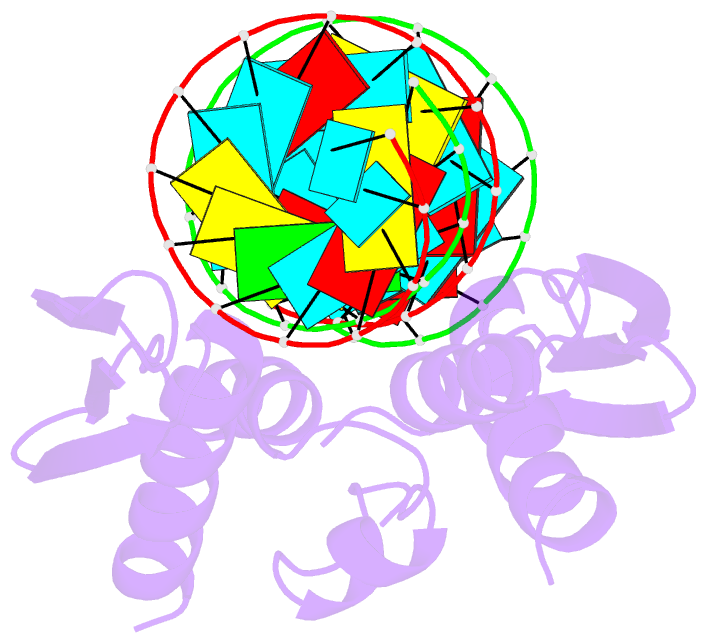
- Structure of trbp dsrbd 1 and 2 in complex with a 19 bp sirna (complex b)
- Reference
- Masliah G, Maris C, Konig SL, Yulikov M, Aeschimann F, Malinowska AL, Mabille J, Weiler J, Holla A, Hunziker J, Meisner-Kober N, Schuler B, Jeschke G, Allain FH (2018): "Structural basis of siRNA recognition by TRBP double-stranded RNA binding domains." EMBO J., 37. doi: 10.15252/embj.201797089.
- Abstract
- The accurate cleavage of pre-micro(mi)RNAs by Dicer and mi/siRNA guide strand selection are important steps in forming the RNA-induced silencing complex (RISC). The role of Dicer binding partner TRBP in these processes remains poorly understood. Here, we solved the solution structure of the two N-terminal dsRNA binding domains (dsRBDs) of TRBP in complex with a functionally asymmetric siRNA using NMR, EPR, and single-molecule spectroscopy. We find that siRNA recognition by the dsRBDs is not sequence-specific but rather depends on the RNA shape. The two dsRBDs can swap their binding sites, giving rise to two equally populated, pseudo-symmetrical complexes, showing that TRBP is not a primary sensor of siRNA asymmetry. Using our structure to model a Dicer-TRBP-siRNA ternary complex, we show that TRBP's dsRBDs and Dicer's RNase III domains bind a canonical 19 base pair siRNA on opposite sides, supporting a mechanism whereby TRBP influences Dicer-mediated cleavage accuracy by binding the dsRNA region of the pre-miRNA during Dicer cleavage.