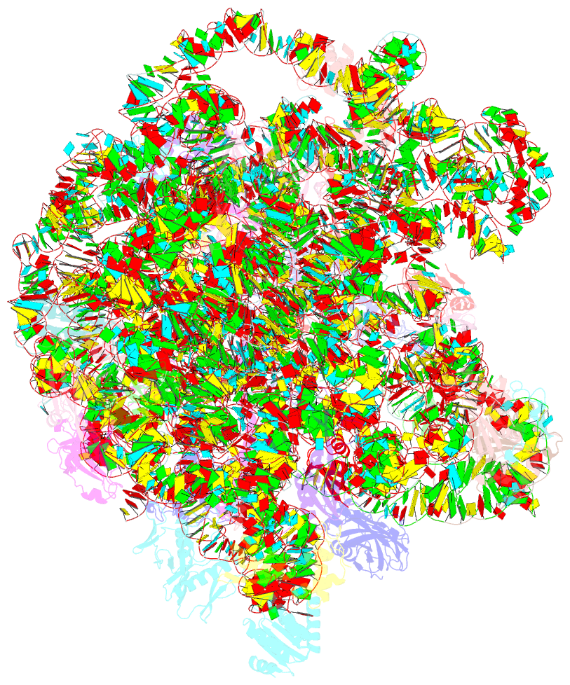
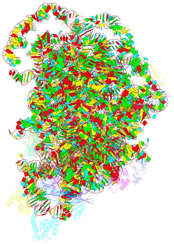
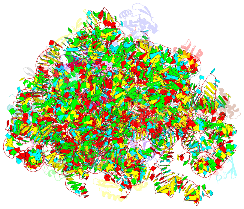
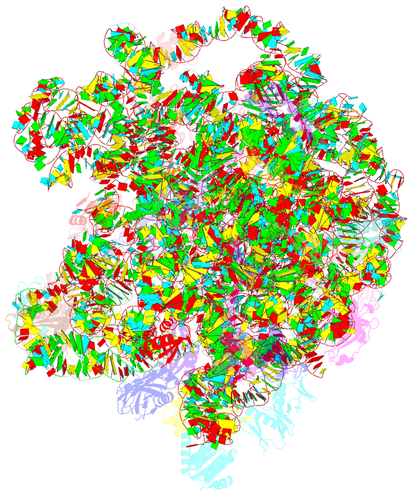
Summary information and primary citation
- PDB-id
- 5o60; SNAP-derived features in text and JSON formats;
DNAproDB
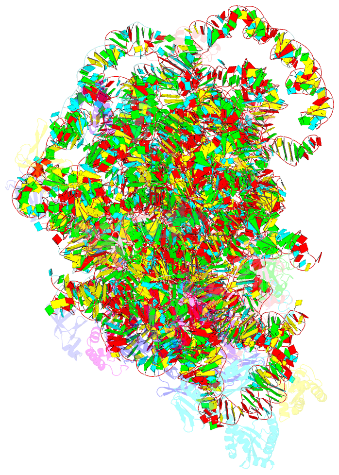
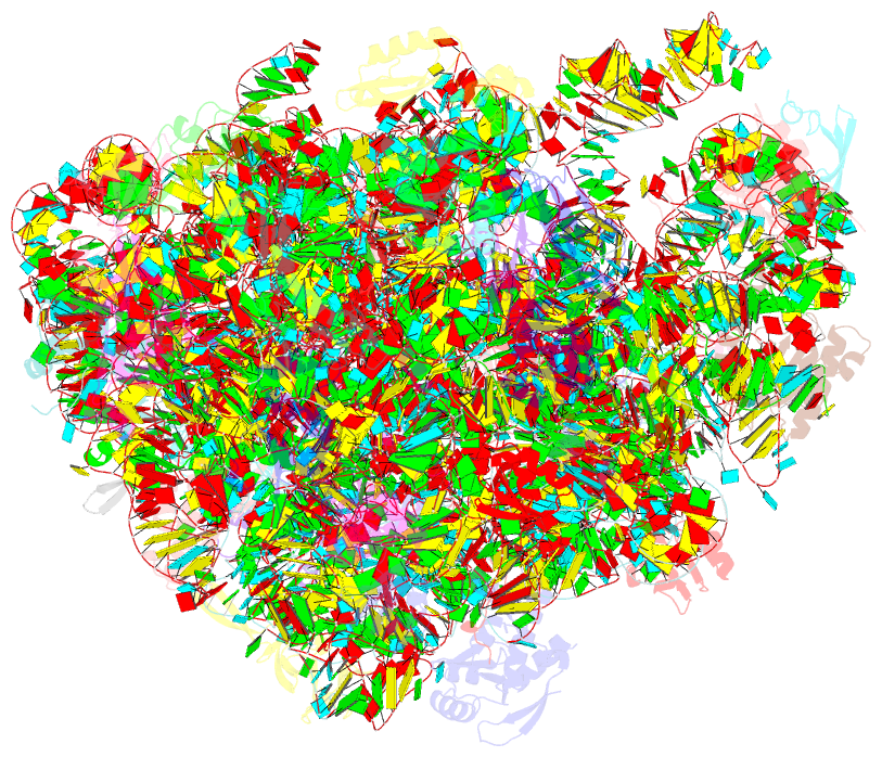
- Class
- ribosome
- Method
- cryo-EM (3.18 Å)
- Summary
- Structure of the 50s large ribosomal subunit from mycobacterium smegmatis
- Reference
- Hentschel J, Burnside C, Mignot I, Leibundgut M, Boehringer D, Ban N (2017): "The Complete Structure of the Mycobacterium smegmatis 70S Ribosome." Cell Rep, 20, 149-160. doi: 10.1016/j.celrep.2017.06.029.
- Abstract
- The ribosome carries out the synthesis of proteins in every living cell. It consequently represents a frontline target in anti-microbial therapy. Tuberculosis ranks among the leading causes of death worldwide, due in large part to the combination of difficult-to-treat latency and antibiotic resistance. Here, we present the 3.3-Å cryo-EM structure of the 70S ribosome of Mycobacterium smegmatis, a close relative to the human pathogen Mycobacterium tuberculosis. The structure reveals two additional ribosomal proteins and localizes them to the vicinity of drug-target sites in both the catalytic center and the decoding site of the ribosome. Furthermore, we visualized actinobacterium-specific rRNA and protein expansions that extensively remodel the ribosomal surface with implications for polysome organization. Our results provide a foundation for understanding the idiosyncrasies of mycobacterial translation and reveal atomic details of the structure that will facilitate the design of anti-tubercular therapeutics.