Summary information and primary citation
- PDB-id
- 5w2m; SNAP-derived features in text and JSON formats;
DNAproDB
- Class
- DNA binding protein
- Method
- X-ray (3.7 Å)
- Summary
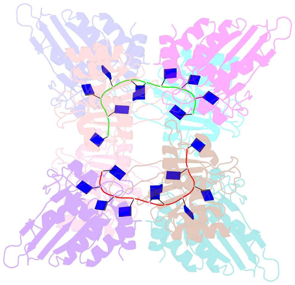
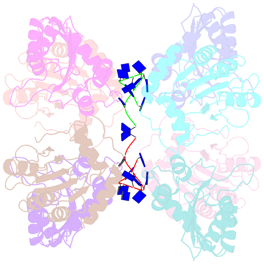
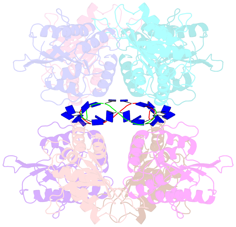
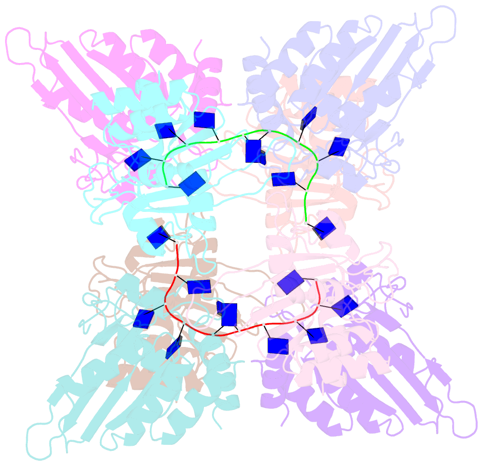
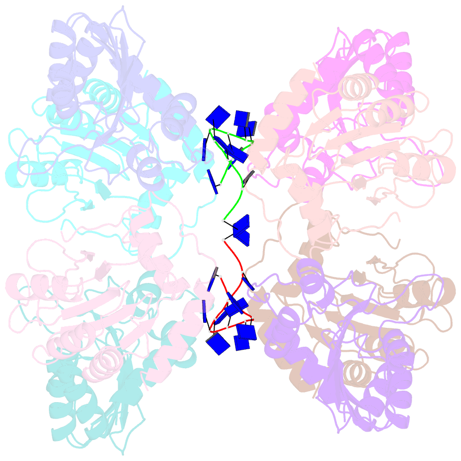
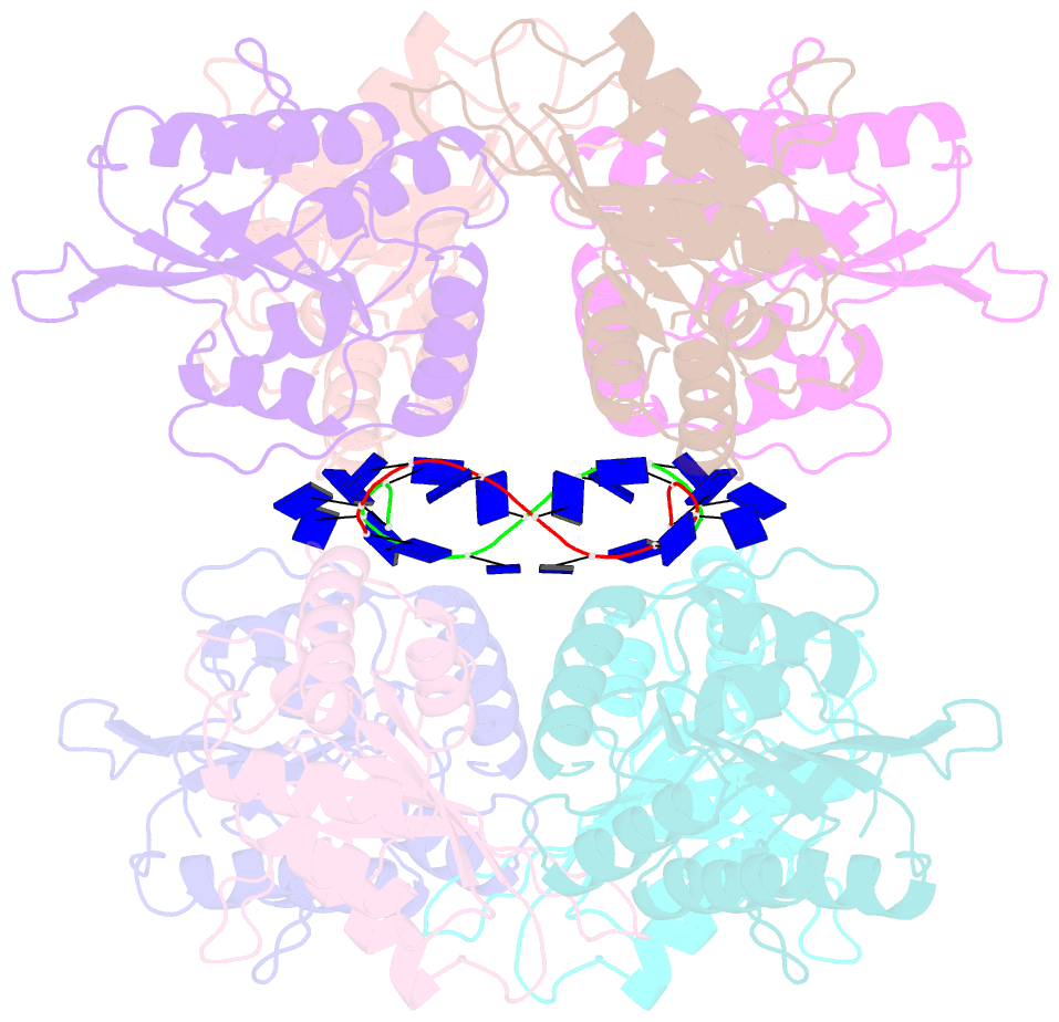
- Apobec3f catalytic domain complex with a single-stranded DNA
- Reference
- Fang Y, Xiao X, Li SX, Wolfe A, Chen XS (2018): "Molecular Interactions of a DNA Modifying Enzyme APOBEC3F Catalytic Domain with a Single-Stranded DNA." J. Mol. Biol., 430, 87-101. doi: 10.1016/j.jmb.2017.11.007.
- Abstract
- The single-stranded DNA (ssDNA) cytidine deaminase APOBEC3F (A3F) deaminates cytosine (C) to uracil (U) and is a known restriction factor of HIV-1. Its C-terminal catalytic domain (CD2) alone is capable of binding single-stranded nucleic acids and is important for deamination. However, little is known about how the CD2 interacts with ssDNA. Here we report a crystal structure of A3F-CD2 in complex with a 10-nucleotide ssDNA composed of poly-thymine, which reveals a novel positively charged nucleic acid binding site distal to the active center that plays a key role in substrate DNA binding and catalytic activity. Lysine and tyrosine residues within this binding site interact with the ssDNA, and mutating these residues dramatically impairs both ssDNA binding and catalytic activity. This binding site is not conserved in APOBEC3G (A3G), which may explain differences in ssDNA-binding characteristics between A3F-CD2 and A3G-CD2. In addition, we observed an alternative Zn-coordination conformation around the active center. These findings reveal the structural relationships between nucleic acid interactions and catalytic activity of A3F.