Summary information and primary citation
- PDB-id
- 5wcu; SNAP-derived features in text and JSON formats;
DNAproDB
- Class
- chromatin binding protein-DNA
- Method
- X-ray (5.53 Å)
- Summary
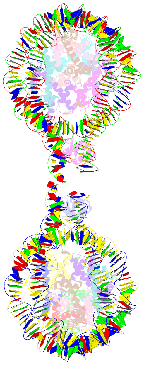
- Crystal structure of 167 bp nucleosome bound to the globular domain of linker histone h5
- Reference
- Zhou BR, Jiang J, Ghirlando R, Norouzi D, Sathish Yadav KN, Feng H, Wang R, Zhang P, Zhurkin V, Bai Y (2018): "Revisit of Reconstituted 30-nm Nucleosome Arrays Reveals an Ensemble of Dynamic Structures." J. Mol. Biol., 430, 3093-3110. doi: 10.1016/j.jmb.2018.06.020.
- Abstract
- It has long been suggested that chromatin may form a fiber with a diameter of ~30 nm that suppresses transcription. Despite nearly four decades of study, the structural nature of the 30-nm chromatin fiber and conclusive evidence of its existence in vivo remain elusive. The key support for the existence of specific 30-nm chromatin fiber structures is based on the determination of the structures of reconstituted nucleosome arrays using X-ray crystallography and single-particle cryo-electron microscopy coupled with glutaraldehyde chemical cross-linking. Here we report the characterization of these nucleosome arrays in solution using analytical ultracentrifugation, NMR, and small-angle X-ray scattering. We found that the physical properties of these nucleosome arrays in solution are not consistent with formation of just a few discrete structures of nucleosome arrays. In addition, we obtained a crystal of the nucleosome in complex with the globular domain of linker histone H5 that shows a new form of nucleosome packing and suggests a plausible alternative compact conformation for nucleosome arrays. Taken together, our results challenge the key evidence for the existence of a limited number of structures of reconstituted nucleosome arrays in solution by revealing that the reconstituted nucleosome arrays are actually best described as an ensemble of various conformations with a zigzagged arrangement of nucleosomes. Our finding has implications for understanding the structure and function of chromatin in vivo.


