Summary information and primary citation
- PDB-id
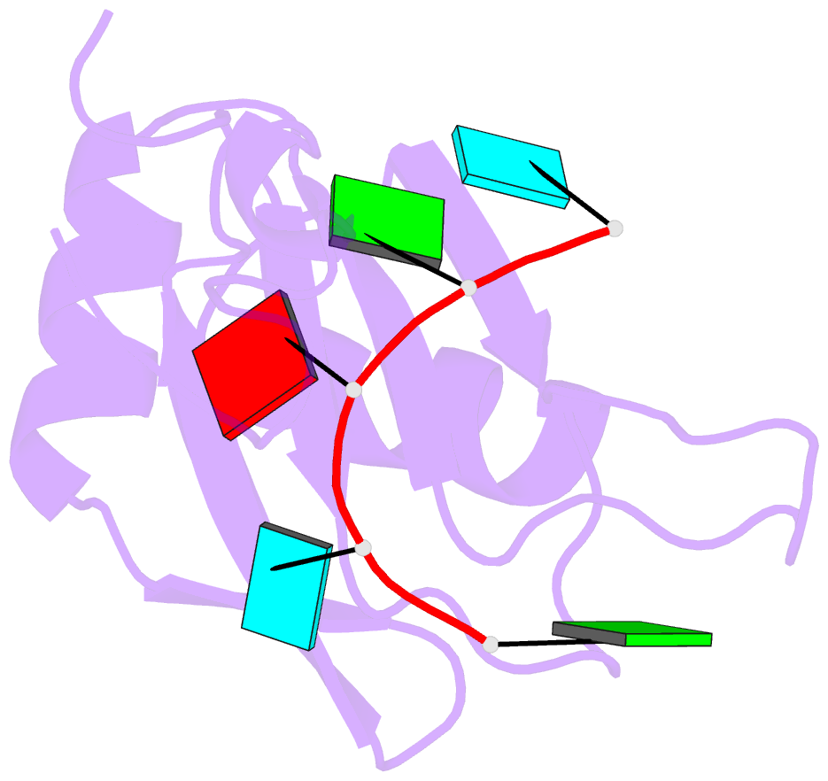
- 5x3z; SNAP-derived features in text and JSON formats;
DNAproDB
- Class
- RNA binding protein-RNA
- Method
- NMR
- Summary
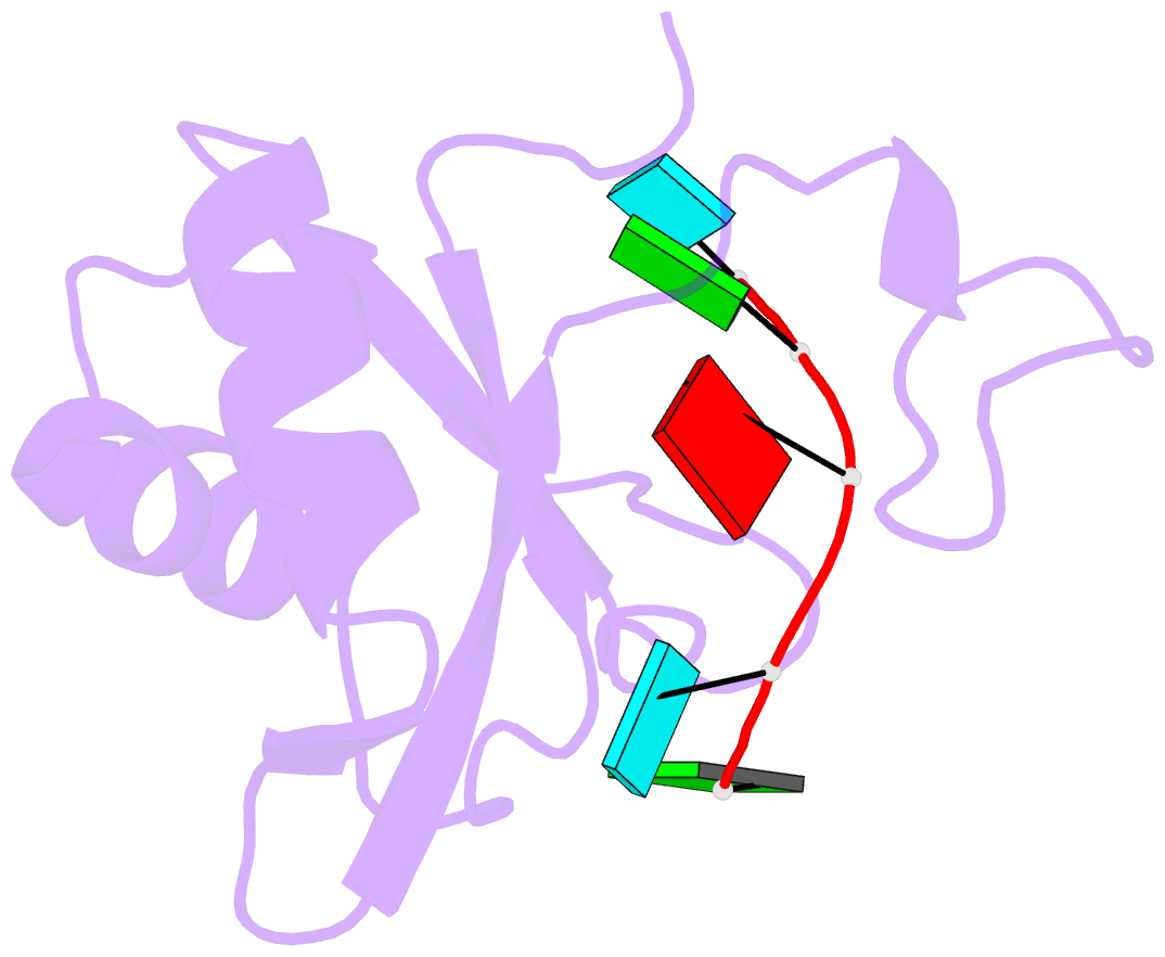
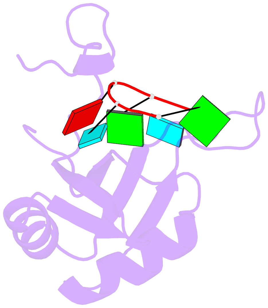
- Solution structure of musashi1 rbd2 in complex with RNA
- Reference
- Iwaoka R, Nagata T, Tsuda K, Imai T, Okano H, Kobayashi N, Katahira M (2017): "Structural Insight into the Recognition of r(UAG) by Musashi-1 RBD2, and Construction of a Model of Musashi-1 RBD1-2 Bound to the Minimum Target RNA." Molecules, 22. doi: 10.3390/molecules22071207.
- Abstract
- Musashi-1 (Msi1) controls the maintenance of stem cells and tumorigenesis through binding to its target mRNAs and subsequent translational regulation. Msi1 has two RNA-binding domains (RBDs), RBD1 and RBD2, which recognize r(GUAG) and r(UAG), respectively. These minimal recognition sequences are connected by variable linkers in the Msi1 target mRNAs, however, the molecular mechanism by which Msi1 recognizes its targets is not yet understood. We previously determined the solution structure of the Msi1 RBD1:r(GUAGU) complex. Here, we determined the first structure of the RBD2:r(GUAGU) complex. The structure revealed that the central trinucleotide, r(UAG), is specifically recognized by the intermolecular hydrogen-bonding and aromatic stacking interactions. Importantly, the C-terminal region, which is disordered in the free form, took a certain conformation, resembling a helix. The observation of chemical shift perturbation and intermolecular NOEs, together with increases in the heteronuclear steady-state {¹H}-15N NOE values on complex formation, indicated the involvement of the C-terminal region in RNA binding. On the basis of the two complex structures, we built a structural model of consecutive RBDs with r(UAGGUAG) containing both minimal recognition sequences, which resulted in no steric hindrance. The model suggests recognition of variable lengths (n) of the linker up to n = 50 may be possible.