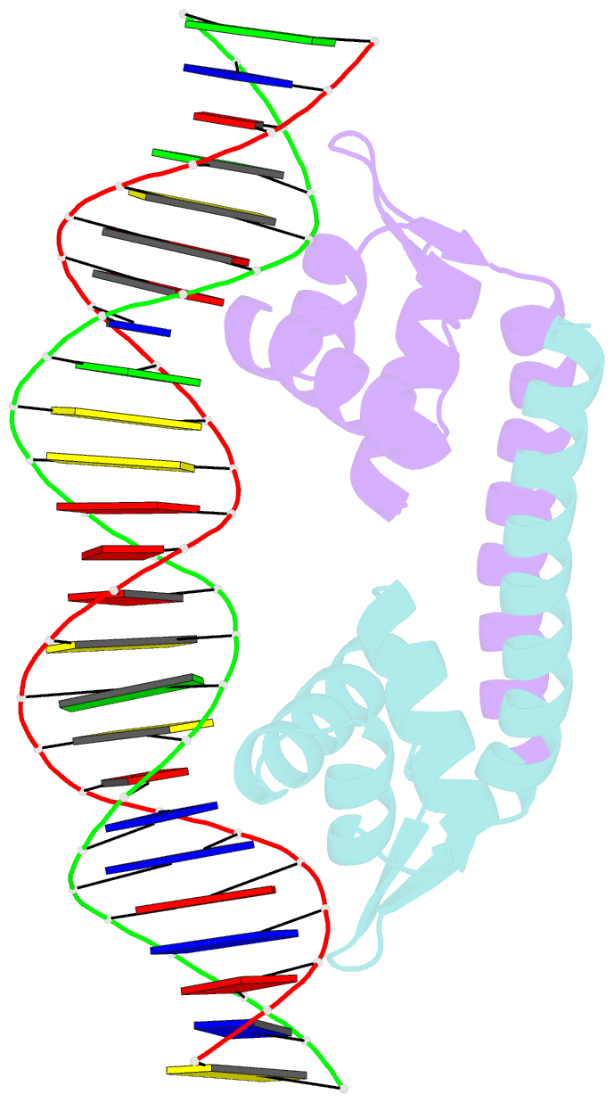
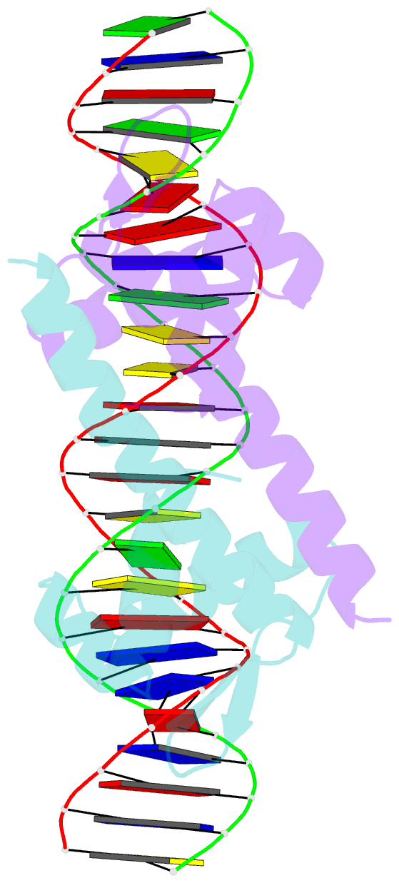
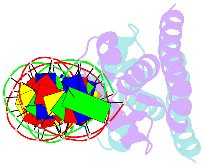
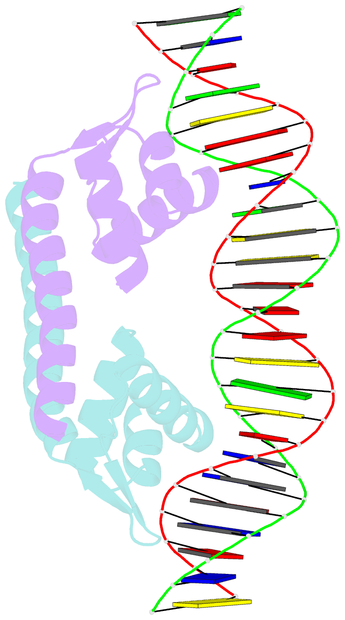
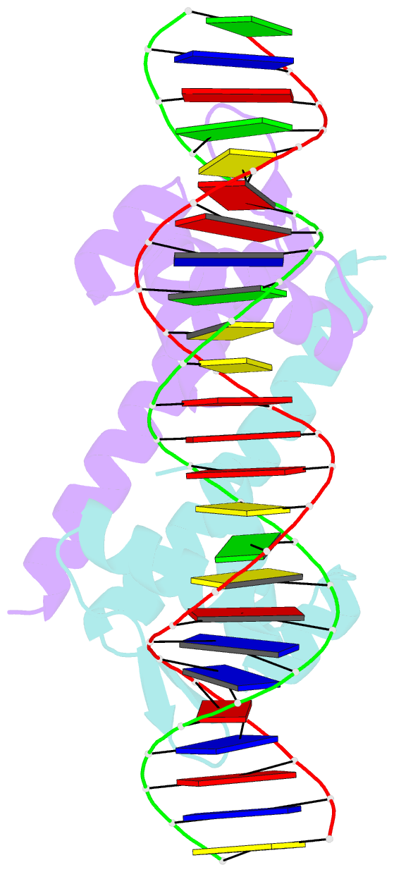
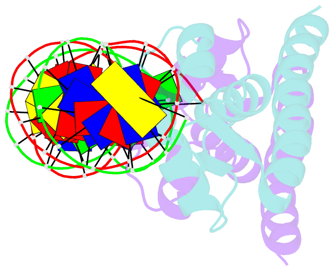
Summary information and primary citation

PDB-id
5xxp; SNAP-derived features in text and JSON formats; DNAproDB
Class
DNA binding protein
Method
X-ray (2.55 Å)
Summary
Crystal structure of cbnr_dbd-DNA complex
Reference
Koentjoro MP, Adachi N, Senda M, Ogawa N, Senda T (2018): "Crystal structure of the DNA-binding domain of the LysR-type transcriptional regulator CbnR in complex with a DNA fragment of the recognition-binding site in the promoter region." FEBS J., 285, 977-989. doi: 10.1111/febs.14380.
Abstract
LysR-type transcriptional regulators (LTTRs) are among the most abundant transcriptional regulators in bacteria. CbnR is an LTTR derived from Cupriavidus necator (formerly Alcaligenes eutrophus or Ralstonia eutropha) NH9 and is involved in transcriptional activation of the cbnABCD genes encoding chlorocatechol degradative enzymes. CbnR interacts with a cbnA promoter region of approximately 60 bp in length that contains the recognition-binding site (RBS) and activation-binding site (ABS). Upon inducer binding, CbnR seems to undergo conformational changes, leading to the activation of the transcription. Since the interaction of an LTTR with RBS is considered to be the first step of the transcriptional activation, the CbnR-RBS interaction is responsible for the selectivity of the promoter to be activated. To understand the sequence selectivity of CbnR, we determined the crystal structure of the DNA-binding domain of CbnR in complex with RBS of the cbnA promoter at 2.55 Å resolution. The crystal structure revealed details of the interactions between the DNA-binding domain and the promoter DNA. A comparison with the previously reported crystal structure of the DNA-binding domain of BenM in complex with its cognate RBS showed several differences in the DNA interactions, despite the structural similarity between CbnR and BenM. These differences explain the observed promoter sequence selectivity between CbnR and BenM. Particularly, the difference between Thr33 in CbnR and Ser33 in BenM appears to affect the conformations of neighboring residues, leading to the selective interactions with DNA.

Cartoon-block schematics in six views (download the tarball)

PyMOL session file Download PDB file View in 3Dmol.js