Summary information and primary citation
- PDB-id
- 5zcw; SNAP-derived features in text and JSON formats;
DNAproDB
- Class
- DNA binding protein-DNA
- Method
- X-ray (2.7 Å)
- Summary
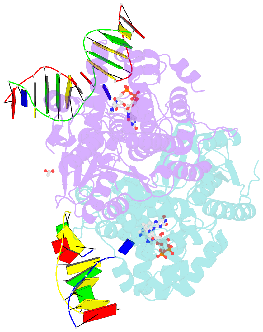
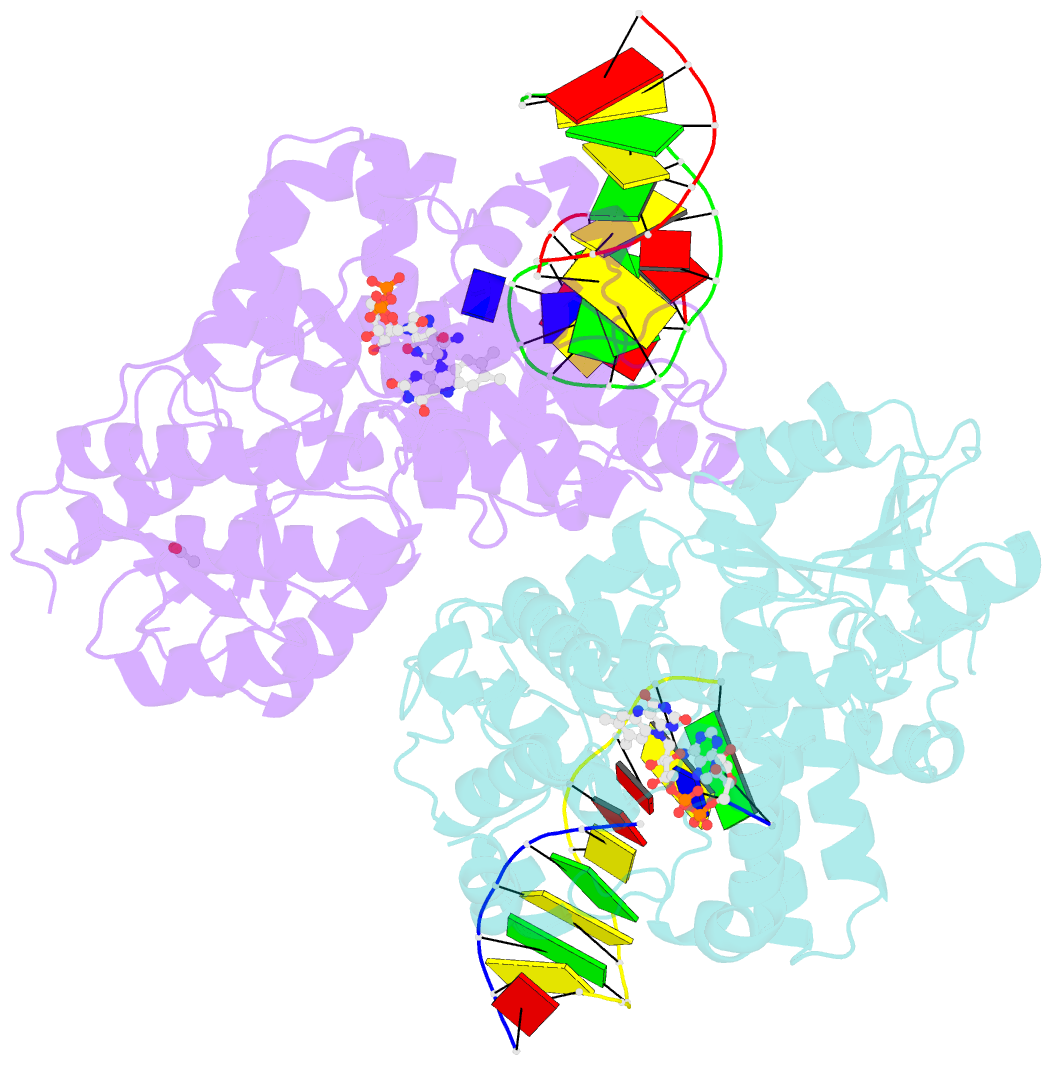
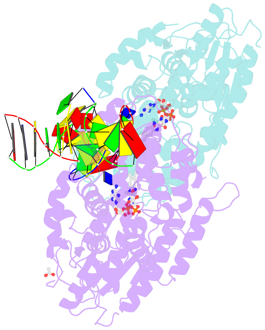
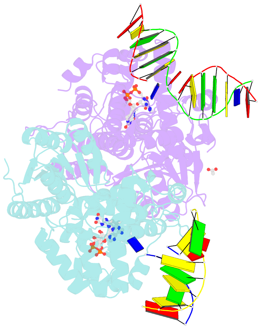
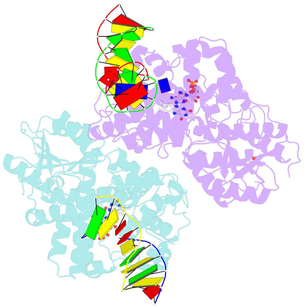
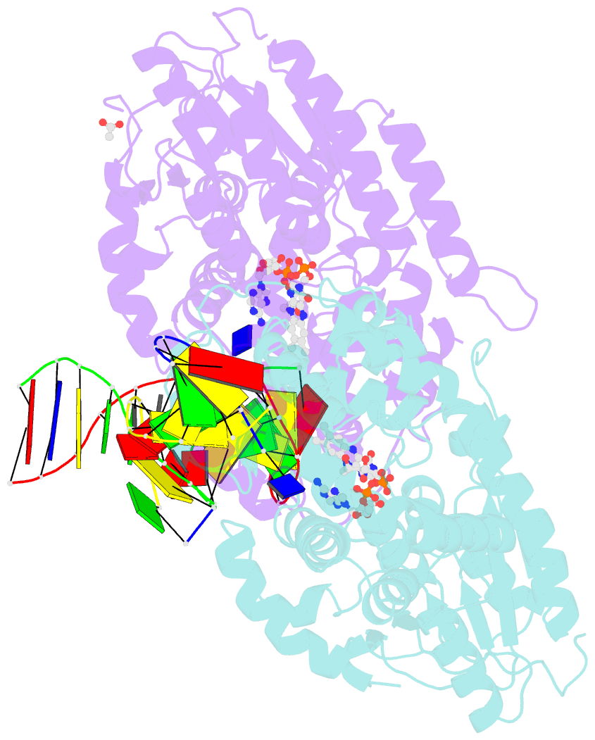
- Structure of the methanosarcina mazei class ii cpd-photolyase in complex with intact, phosphodiester linked, cpd-lesion
- Reference
- Maestre-Reyna M, Yamamoto J, Huang WC, Tsai MD, Essen LO, Bessho Y (2018): "Twist and turn: a revised structural view on the unpaired bubble of class II CPD photolyase in complex with damaged DNA." IUCrJ, 5, 608-618. doi: 10.1107/S205225251800996X.
- Abstract
- Cyclobutane pyrimidine dimer (CPD) photolyases harness the energy of blue light to repair UV-induced DNA CPDs. Upon binding, CPD photolyases cause the photodamage to flip out of the duplex DNA and into the catalytic site of the enzyme. This process, called base-flipping, induces a kink in the DNA, as well as an unpaired bubble, which are stabilized by a network of protein-nucleic acid interactions. Previously, several co-crystal structures have been reported in which the binding mode of CPD photolyases has been studied in detail. However, in all cases the internucleoside linkage of the photodamage site was a chemically synthesized formacetal analogue and not the natural phosphodiester. Here, the first crystal structure and conformational analysis via molecular-dynamics simulations of a class II CPD photolyase in complex with photodamaged DNA that contains a natural cyclobutane pyrimidine dimer with an intra-lesion phosphodiester linkage are presented. It is concluded that a highly conserved bubble-intruding region (BIR) mediates stabilization of the open form of CPD DNA when complexed with class II CPD photolyases.