Summary information and primary citation
- PDB-id
- 6bk8; SNAP-derived features in text and JSON formats;
DNAproDB
- Class
- RNA binding protein
- Method
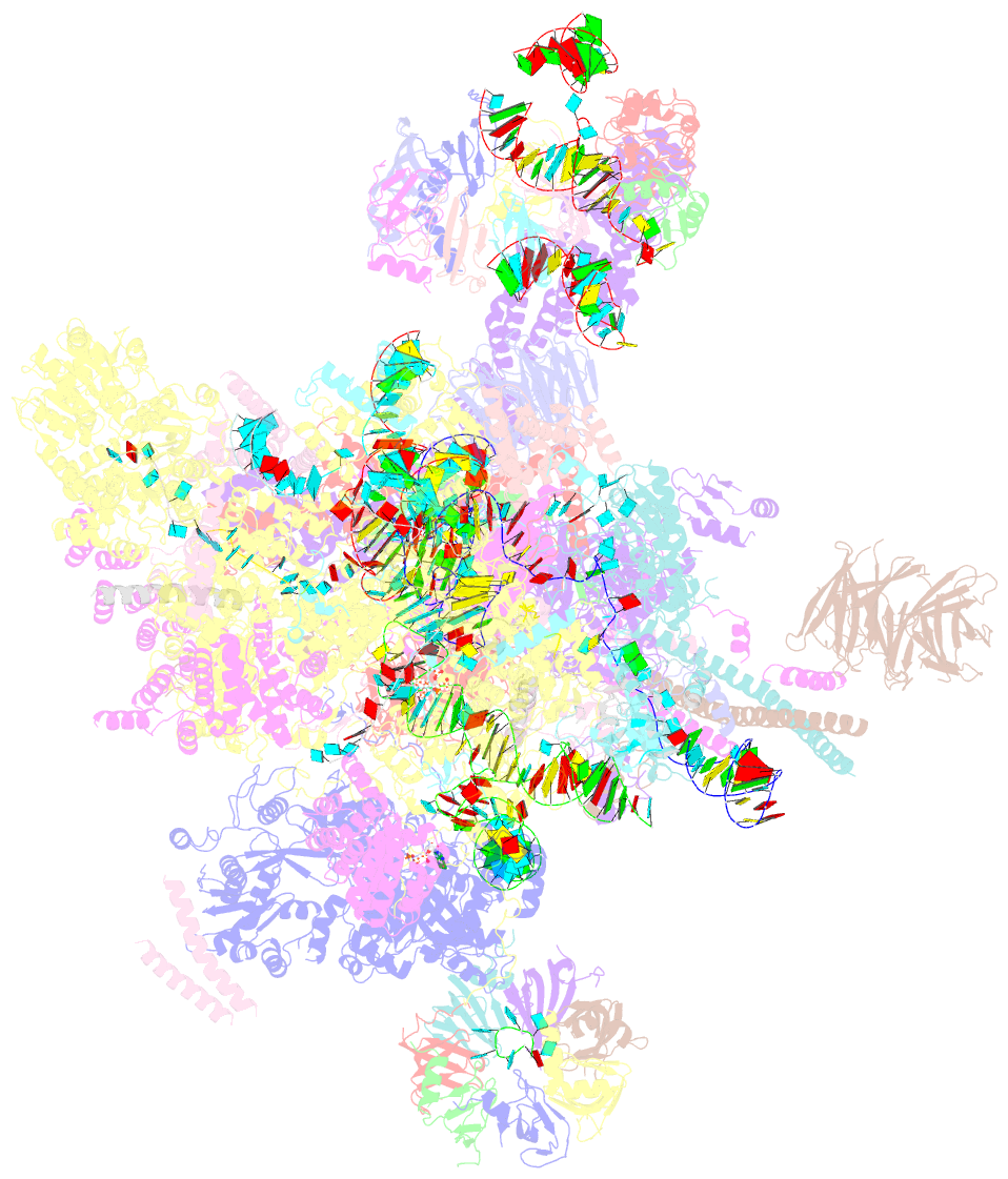
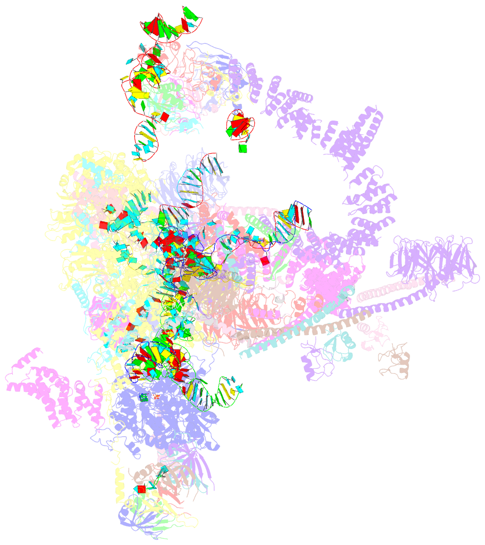
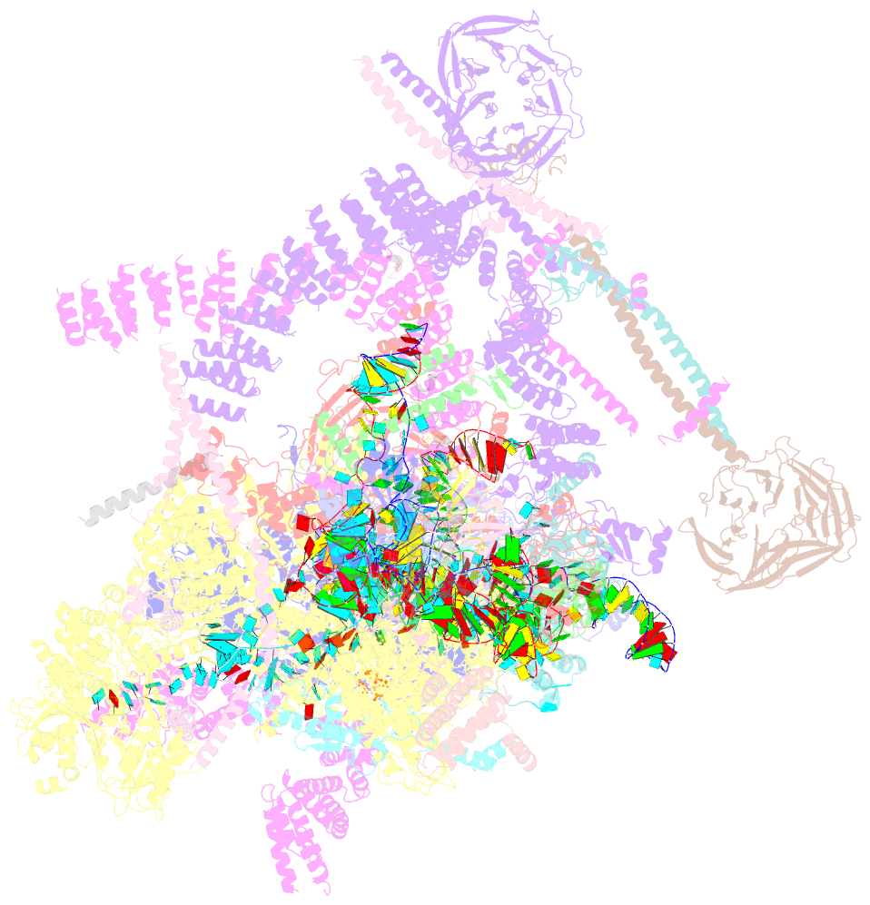
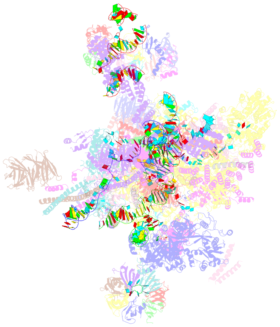
- cryo-EM (3.3 Å)
- Summary
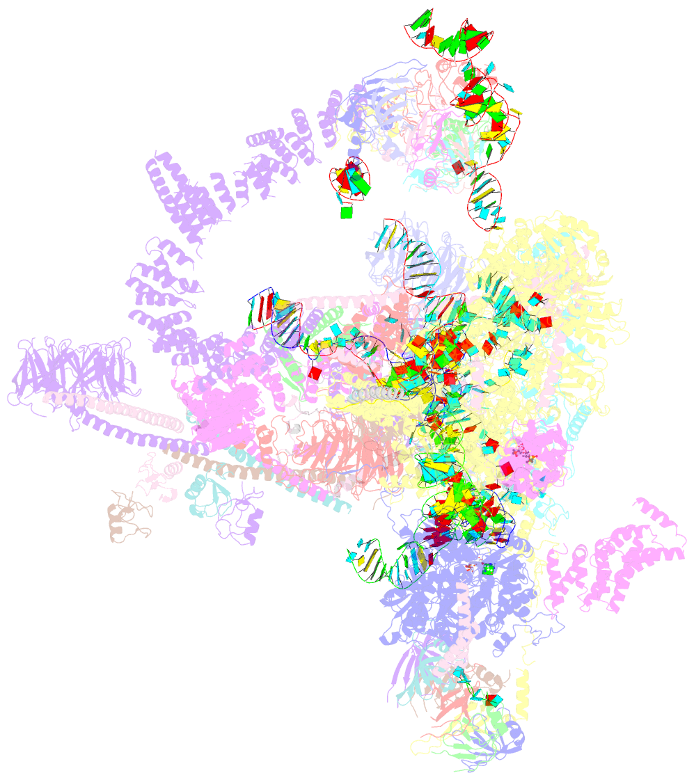
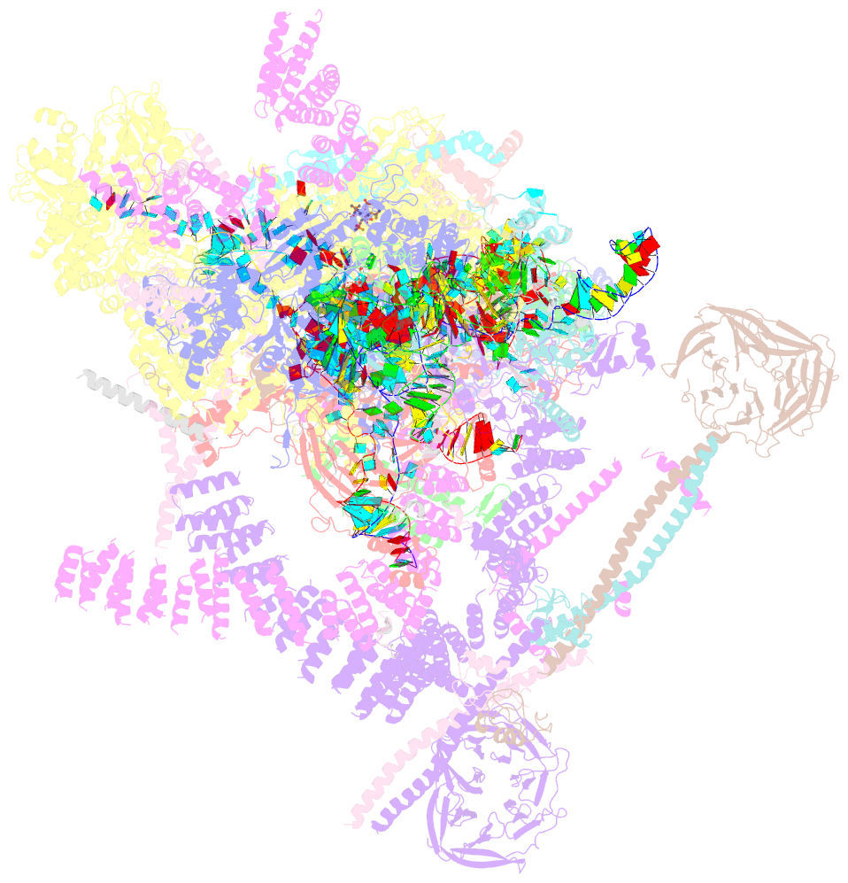
- S. cerevisiae spliceosomal post-catalytic p complex
- Reference
- Liu S, Li X, Zhang L, Jiang J, Hill RC, Cui Y, Hansen KC, Zhou ZH, Zhao R (2017): "Structure of the yeast spliceosomal postcatalytic P complex." Science, 358, 1278-1283. doi: 10.1126/science.aar3462.
- Abstract
- The spliceosome undergoes dramatic changes in a splicing cycle. Structures of B, Bact, C, C*, and intron lariat spliceosome complexes revealed mechanisms of 5'-splice site (ss) recognition, branching, and intron release, but lacked information on 3'-ss recognition, exon ligation, and exon release. Here we report a cryo-electron microscopy structure of the postcatalytic P complex at 3.3-angstrom resolution, revealing that the 3' ss is mainly recognized through non-Watson-Crick base pairing with the 5' ss and branch point. Furthermore, one or more unidentified proteins become stably associated with the P complex, securing the 3' exon and potentially regulating activity of the helicase Prp22. Prp22 binds nucleotides 15 to 21 in the 3' exon, enabling it to pull the intron-exon or ligated exons in a 3' to 5' direction to achieve 3'-ss proofreading or exon release, respectively.