Summary information and primary citation
- PDB-id
- 6evj; SNAP-derived features in text and JSON formats;
DNAproDB
- Class
- viral protein
- Method
- X-ray (3.9 Å)
- Summary
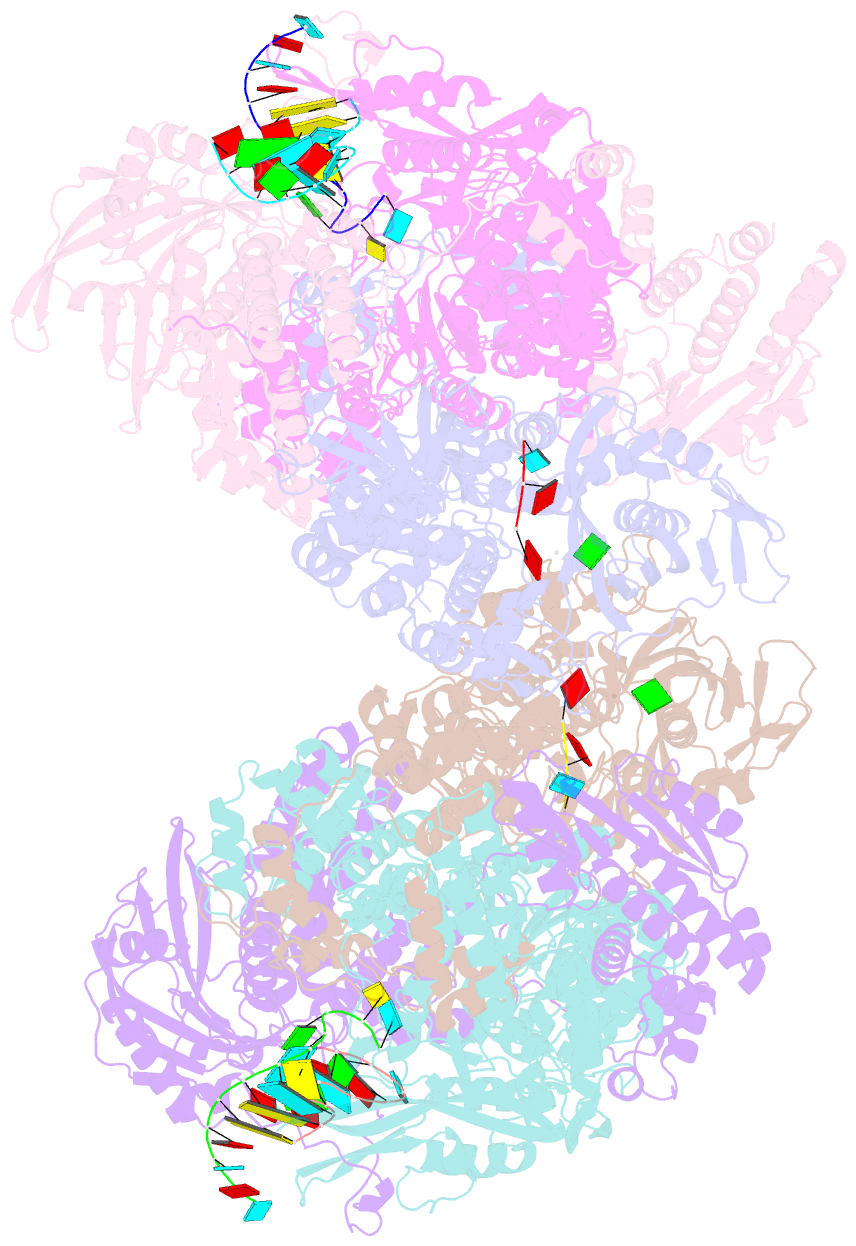
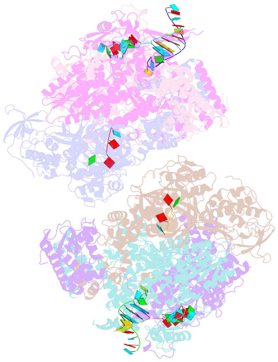
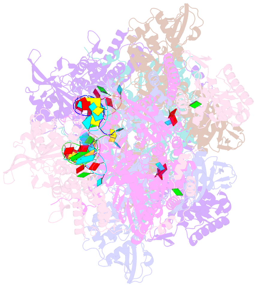
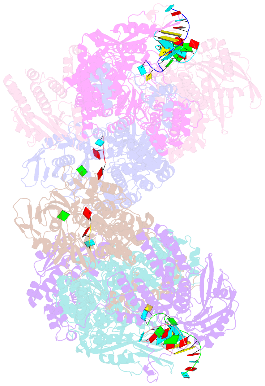
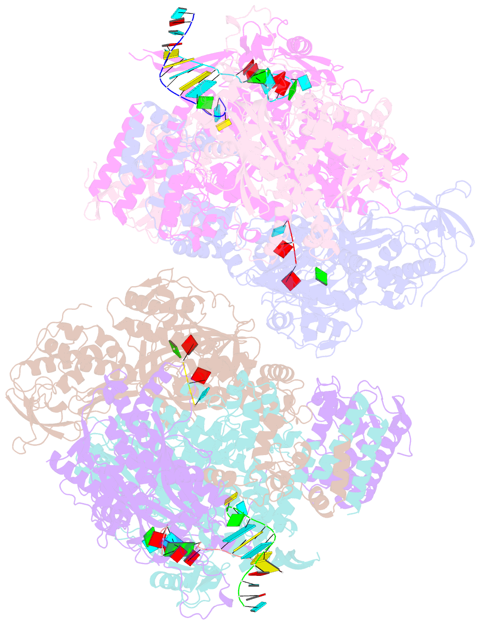
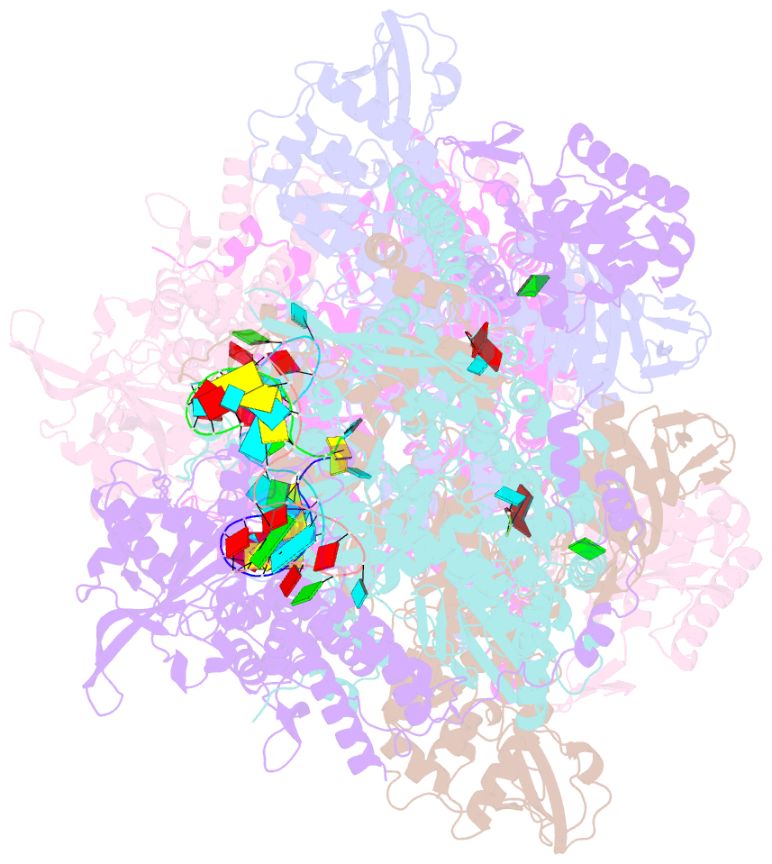
- Crystal structure of bat influenza a-h17n10 polymerase with viral RNA promoter and capped RNA primer
- Reference
- Pflug A, Gaudon S, Resa-Infante P, Lethier M, Reich S, Schulze WM, Cusack S (2018): "Capped RNA primer binding to influenza polymerase and implications for the mechanism of cap-binding inhibitors." Nucleic Acids Res., 46, 956-971. doi: 10.1093/nar/gkx1210.
- Abstract
- Influenza polymerase uses short capped primers snatched from nascent Pol II transcripts to initiate transcription of viral mRNAs. Here we describe crystal structures of influenza A and B polymerase bound to a capped primer in a configuration consistent with transcription initiation ('priming state') and show by functional assays that conserved residues from both the PB2 midlink and cap-binding domains are important for positioning the capped RNA. In particular, mutation of PB2 Arg264, which interacts with the triphosphate linkage in the cap, significantly and specifically decreases cap-dependent transcription. We also compare the configuration of the midlink and cap-binding domains in the priming state with their very different relative arrangement (called the 'apo' state) in structures where the potent cap-binding inhibitor VX-787, or a close analogue, is bound. In the 'apo' state the inhibitor makes additional interactions to the midlink domain that increases its affinity beyond that to the cap-binding domain alone. The comparison suggests that the mechanism of resistance of certain mutations that allow virus to escape from VX-787, notably PB2 N510T, can only be rationalized if VX-787 has a dual mode of action, direct inhibition of capped RNA binding as well as stabilization of the transcriptionally inactive 'apo' state.