Summary information and primary citation
- PDB-id
- 6htu; SNAP-derived features in text and JSON formats;
DNAproDB
- Class
- RNA binding protein
- Method
- X-ray (2.888 Å)
- Summary
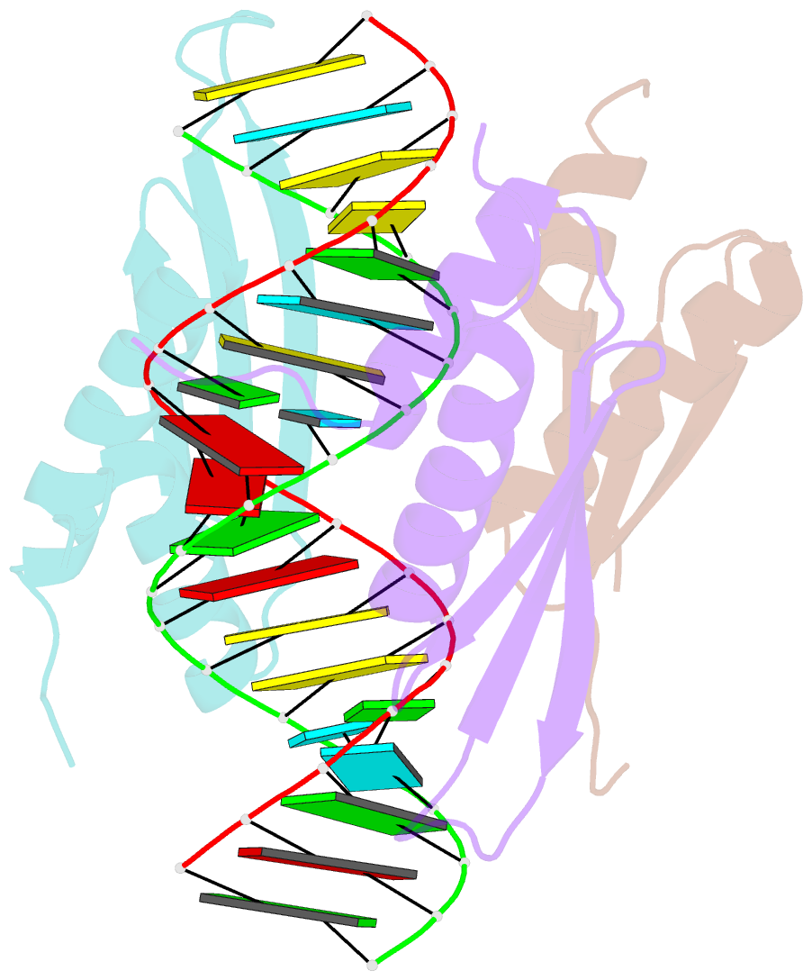
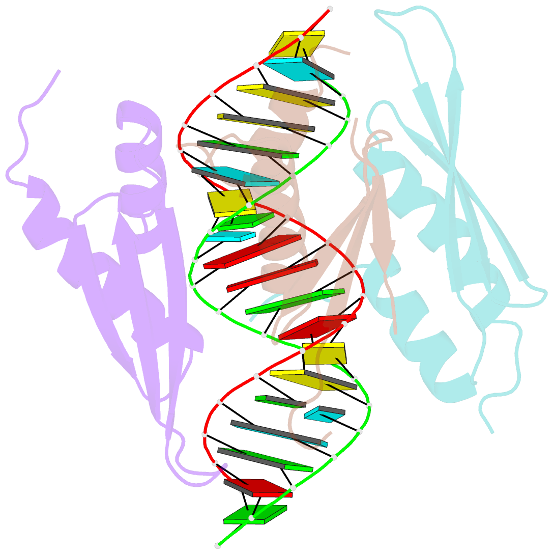
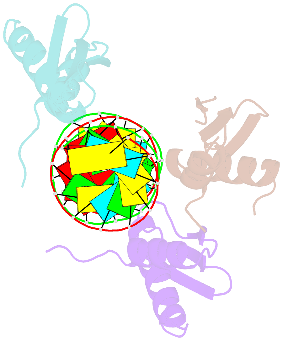
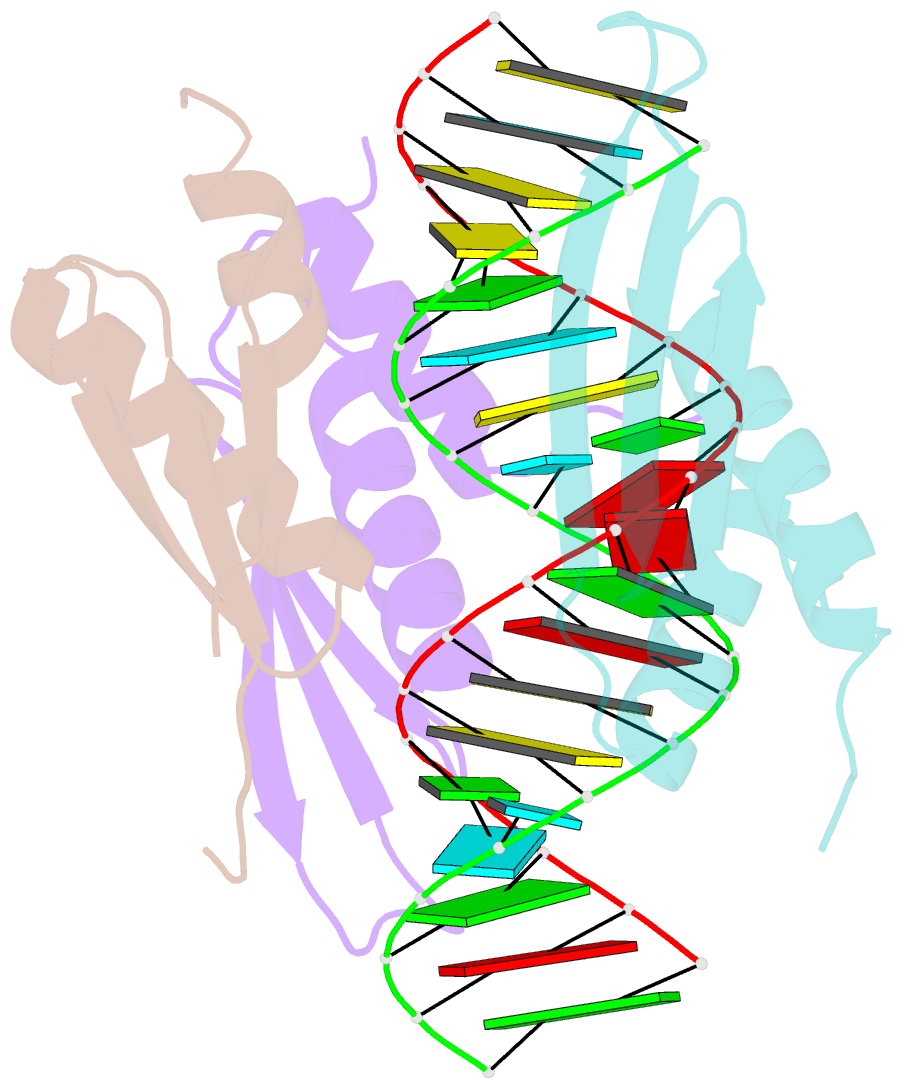
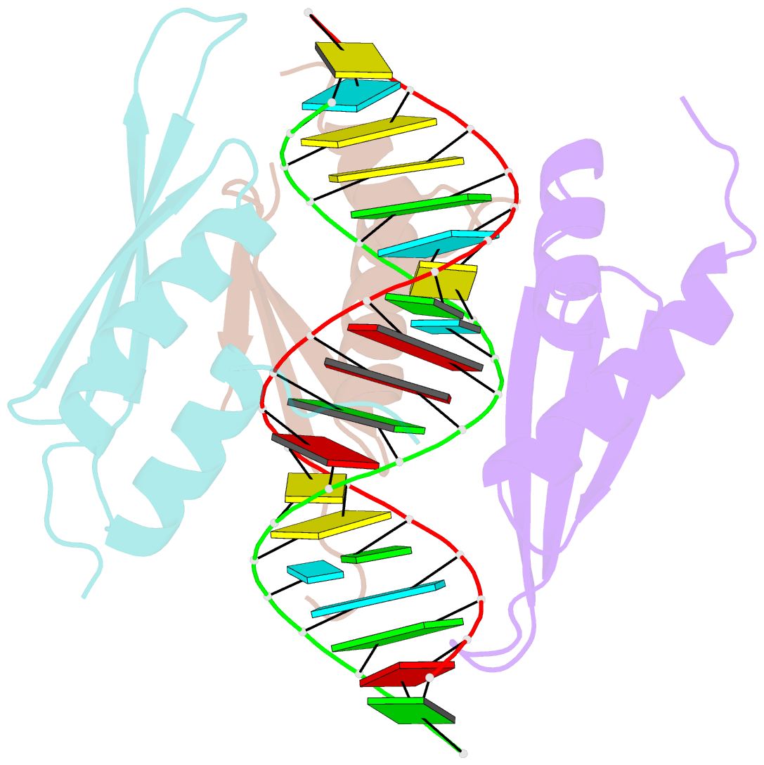
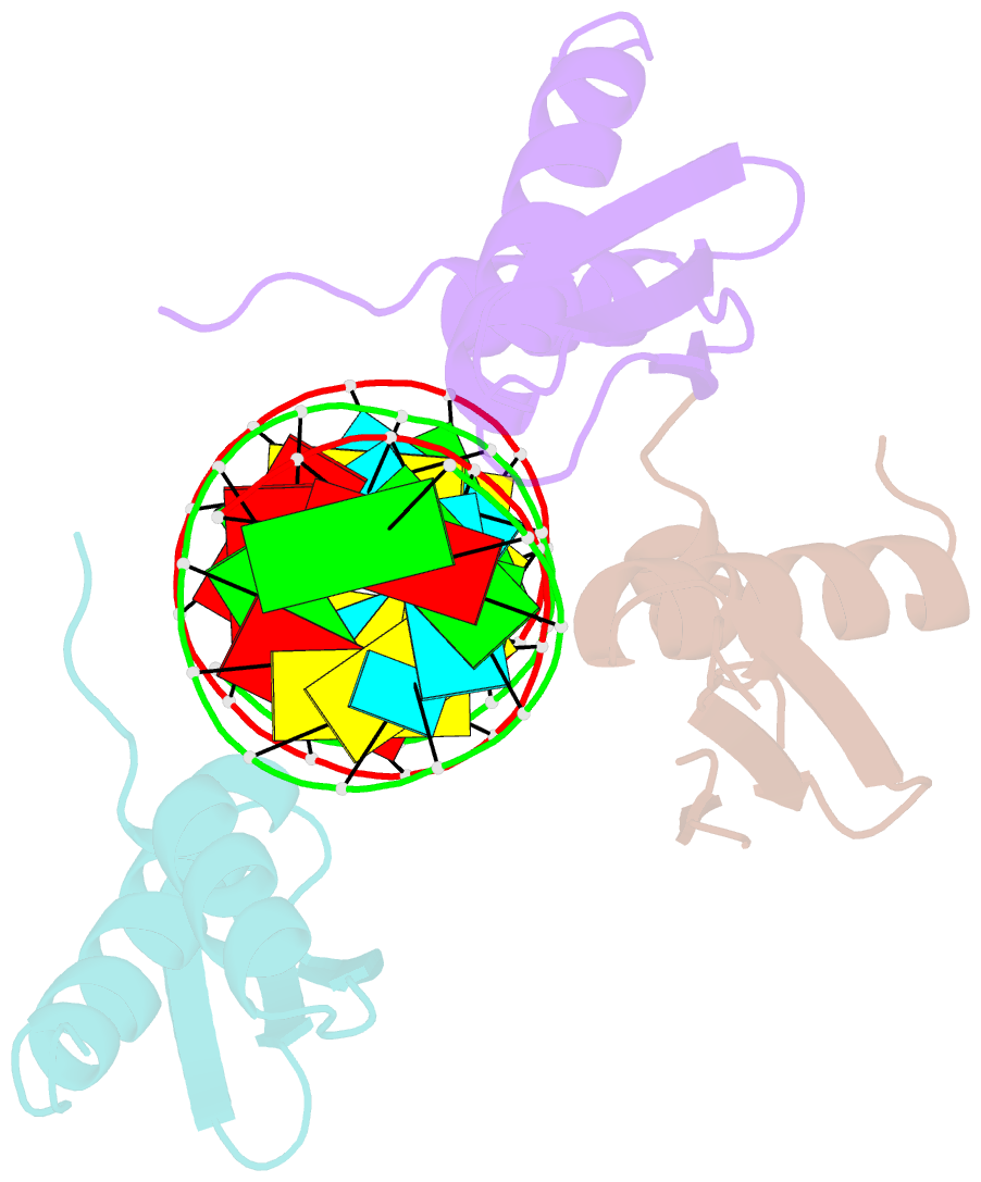
- Structure of hstau1 dsrbd3-4 in complex with arf1 RNA
- Reference
- Lazzaretti D, Bandholz-Cajamarca L, Emmerich C, Schaaf K, Basquin C, Irion U, Bono F (2018): "The crystal structure of Staufen1 in complex with a physiological RNA sheds light on substrate selectivity." Life Sci Alliance, 1, e201800187. doi: 10.26508/lsa.201800187.
- Abstract
- During mRNA localization, RNA-binding proteins interact with specific structured mRNA localization motifs. Although several such motifs have been identified, we have limited structural information on how these interact with RNA-binding proteins. Staufen proteins bind structured mRNA motifs through dsRNA-binding domains (dsRBD) and are involved in mRNA localization in Drosophila and mammals. We solved the structure of two dsRBDs of human Staufen1 in complex with a physiological dsRNA sequence. We identified interactions between the dsRBDs and the RNA sugar-phosphate backbone and direct contacts of conserved Staufen residues to RNA bases. Mutating residues mediating nonspecific backbone interactions only affected Staufen function in Drosophila when in vitro binding was severely reduced. Conversely, residues involved in base-directed interactions were required in vivo even when they minimally affected in vitro binding. Our work revealed that Staufen can read sequence features in the minor groove of dsRNA and suggests that these influence target selection in vivo.