Summary information and primary citation
- PDB-id
- 6ij2; SNAP-derived features in text and JSON formats;
DNAproDB
- Class
- hydrolase
- Method
- X-ray (1.7 Å)
- Summary
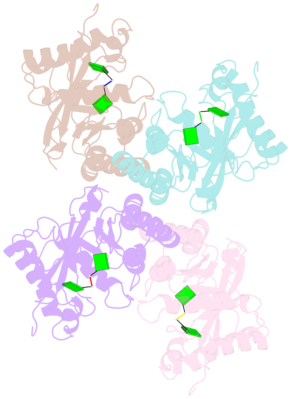
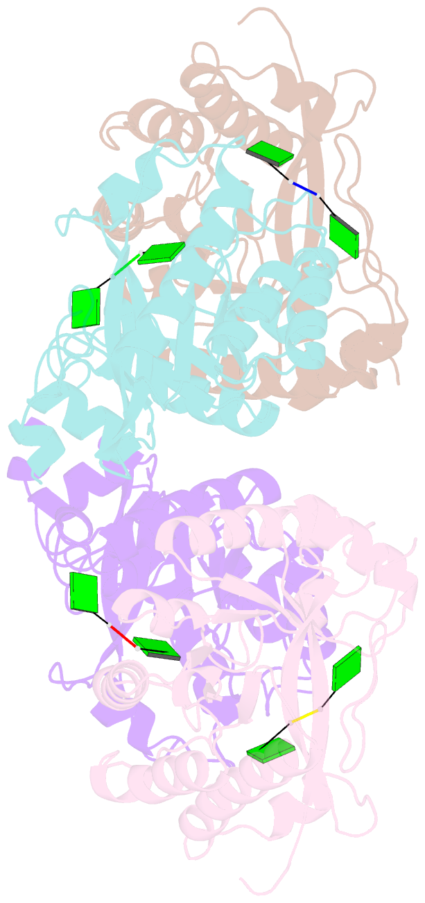
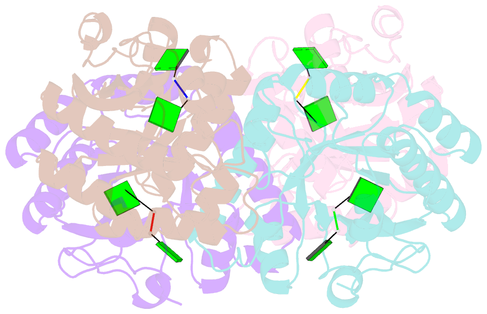
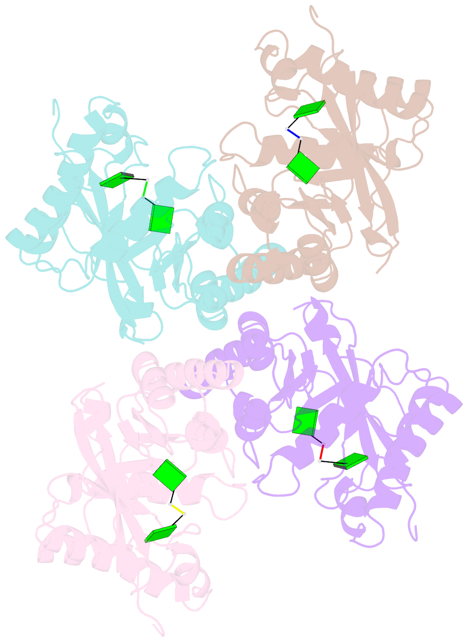
- Crystal structure of a standalone versatile eal protein from vibrio cholerae o395 - 5'-pgpg bound form
- Reference
- Yadav M, Pal K, Sen U (2019): "Structures of c-di-GMP/cGAMP degrading phosphodiesterase VcEAL: identification of a novel conformational switch and its implication." Biochem.J., 476, 3333-3353. doi: 10.1042/BCJ20190399.
- Abstract
- Cyclic dinucleotides (CDNs) have emerged as the central molecules that aid bacteria to adapt and thrive in changing environmental conditions. Therefore, tight regulation of intracellular CDN concentration by counteracting the action of dinucleotide cyclases and phosphodiesterases (PDEs) is critical. Here, we demonstrate that a putative stand-alone EAL domain PDE from Vibrio cholerae (VcEAL) is capable to degrade both the second messenger c-di-GMP and hybrid 3'3'-cyclic GMP-AMP (cGAMP). To unveil their degradation mechanism, we have determined high-resolution crystal structures of VcEAL with Ca2+, c-di-GMP-Ca2+, 5'-pGpG-Ca2+ and cGAMP-Ca2+, the latter provides the first structural basis of cGAMP hydrolysis. Structural studies reveal a typical triosephosphate isomerase barrel-fold with substrate c-di-GMP/cGAMP bound in an extended conformation. Highly conserved residues specifically bind the guanine base of c-di-GMP/cGAMP in the G2 site while the semi-conserved nature of residues at the G1 site could act as a specificity determinant. Two metal ions, co-ordinated with six stubbornly conserved residues and two non-bridging scissile phosphate oxygens of c-di-GMP/cGAMP, activate a water molecule for an in-line attack on the phosphodiester bond, supporting two-metal ion-based catalytic mechanism. PDE activity and biofilm assays of several prudently designed mutants collectively demonstrate that VcEAL active site is charge and size optimized. Intriguingly, in VcEAL-5'-pGpG-Ca2+ structure, β5-α5 loop adopts a novel conformation that along with conserved E131 creates a new metal-binding site. This novel conformation along with several subtle changes in the active site designate VcEAL-5'-pGpG-Ca2+ structure quite different from other 5'-pGpG bound structures reported earlier.