Summary information and primary citation
- PDB-id
- 6jr0; SNAP-derived features in text and JSON formats;
DNAproDB
- Class
- gene regulation-DNA
- Method
- X-ray (2.5 Å)
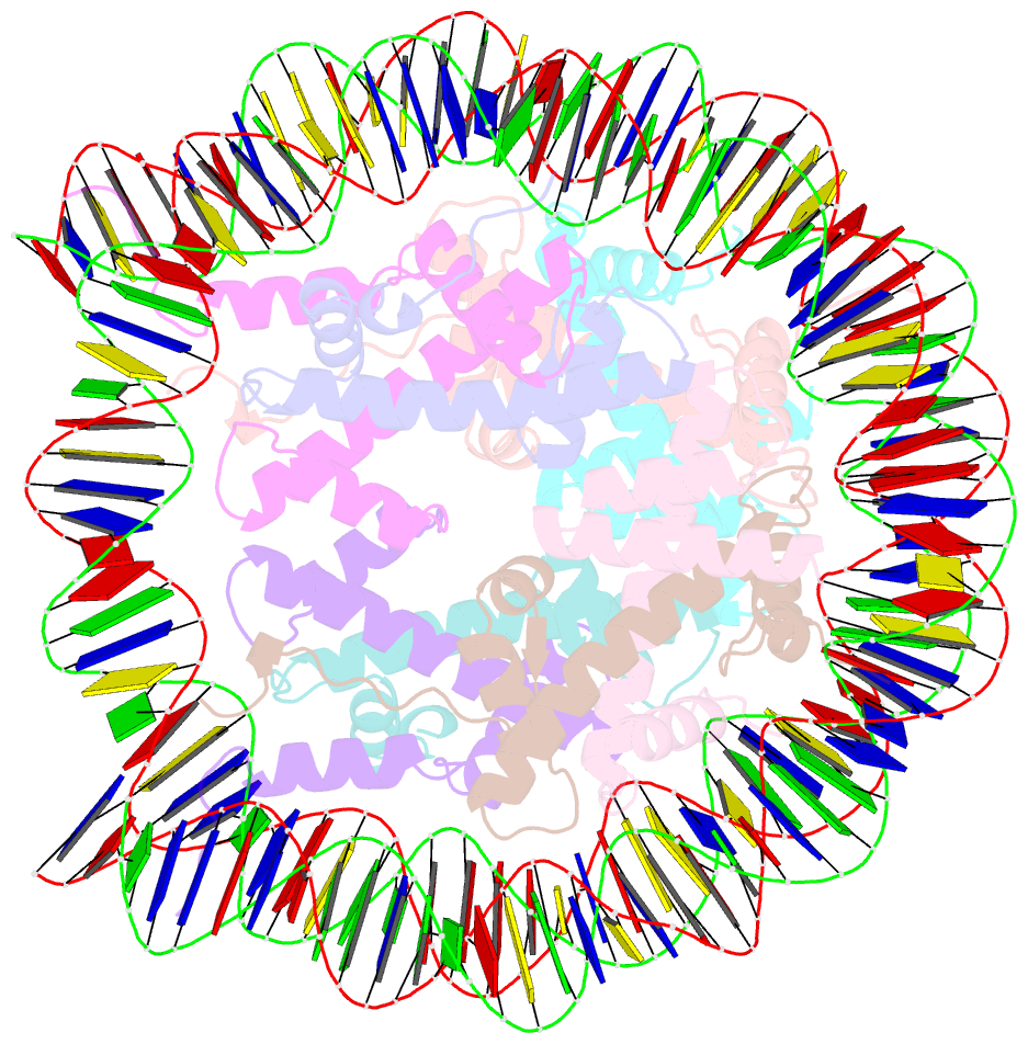
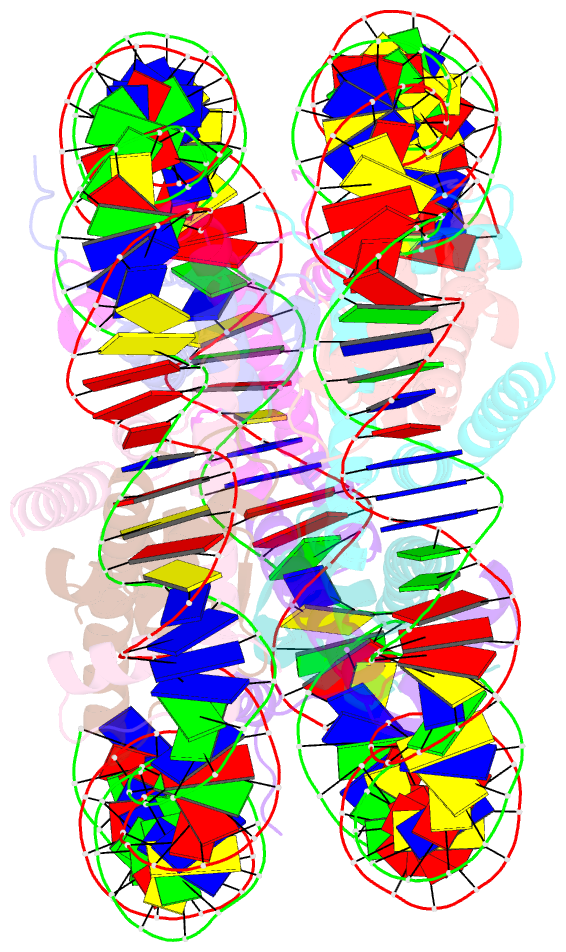
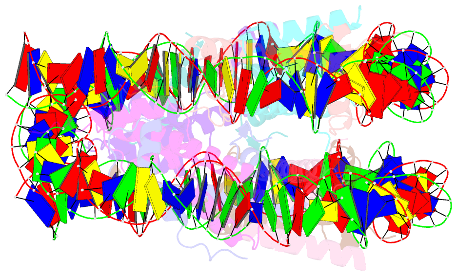
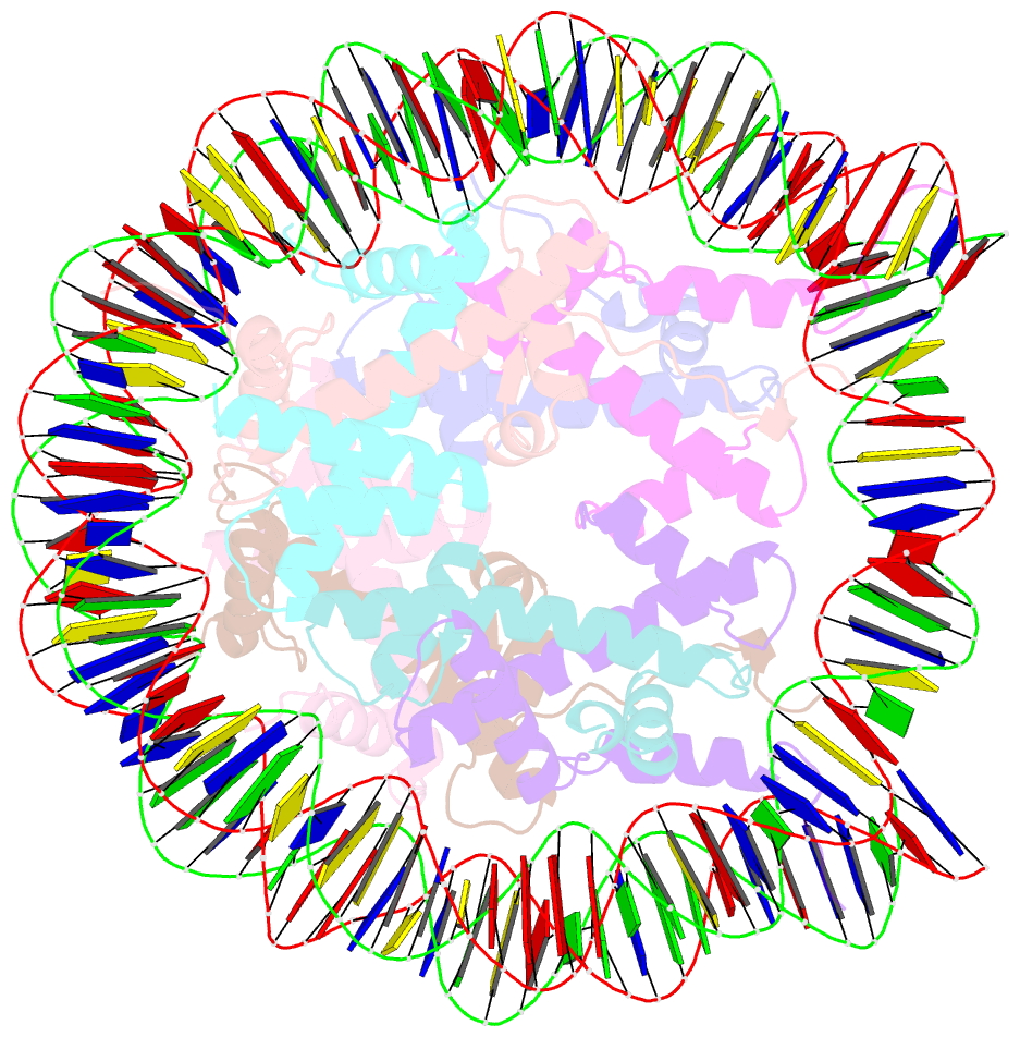
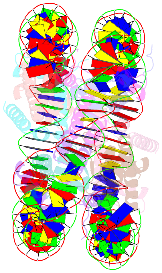
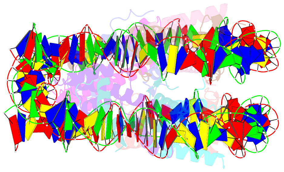
- Summary
- Crystal structure of the human nucleosome phased with 12 selenium atoms
- Reference
- Saotome M, Horikoshi N, Urano K, Kujirai T, Yuzurihara H, Kurumizaka H, Kagawa W (2019): "Structure determination of the nucleosome core particle by selenium SAD phasing." Acta Crystallogr D Struct Biol, 75, 930-936. doi: 10.1107/S2059798319012713.
- Abstract
- The eukaryotic genome is compacted inside the nucleus of the cell in the form called chromatin. The fundamental unit of chromatin is the nucleosome, which contains four types of histones (H3, H4, H2A and H2B) and approximately 150 base pairs of DNA wrapped around the histone complex. The structure of the nucleosome is highly conserved across several eukaryotic species, and molecular replacement has been the primary phasing method used to solve nucleosome structures by X-ray crystallography. However, there is currently no simple, widely applicable experimental phasing method for the nucleosome. In the present study, it is demonstrated that selenomethionine-incorporated histones H3, H2A and H2B can be reconstituted into nucleosomes and crystallized for structural determination. Unexpectedly, it was found that the nucleosome can be phased with a relatively small number of Se atoms. The structures of nucleosome core particles containing 12 and 16 Se atoms were solved by SAD phasing at 2.5 and 2.4 Å resolution, respectively. The present study demonstrates a simple method for determining nucleosome structures by experimental phasing, which may be particularly useful for noncanonical structures that cannot be solved by molecular replacement.