Summary information and primary citation
- PDB-id
- 6n9u; SNAP-derived features in text and JSON formats;
DNAproDB
- Class
- hydrolase,transferase-DNA
- Method
- cryo-EM (3.7 Å)
- Summary
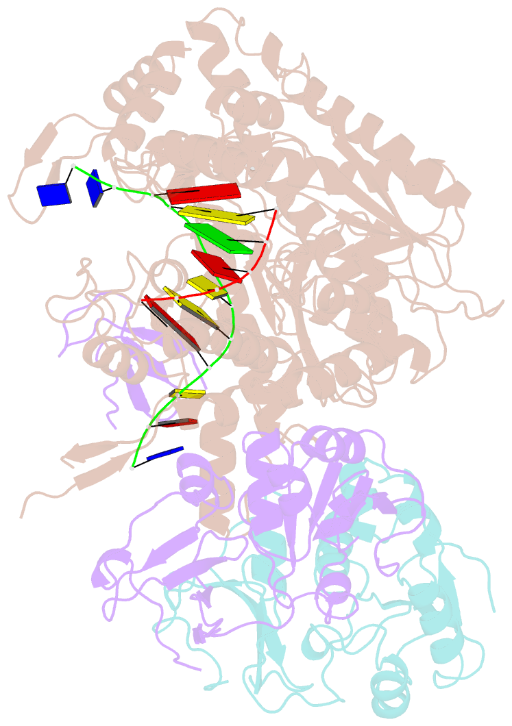
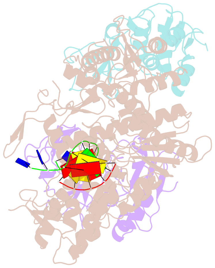
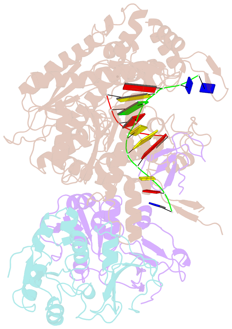
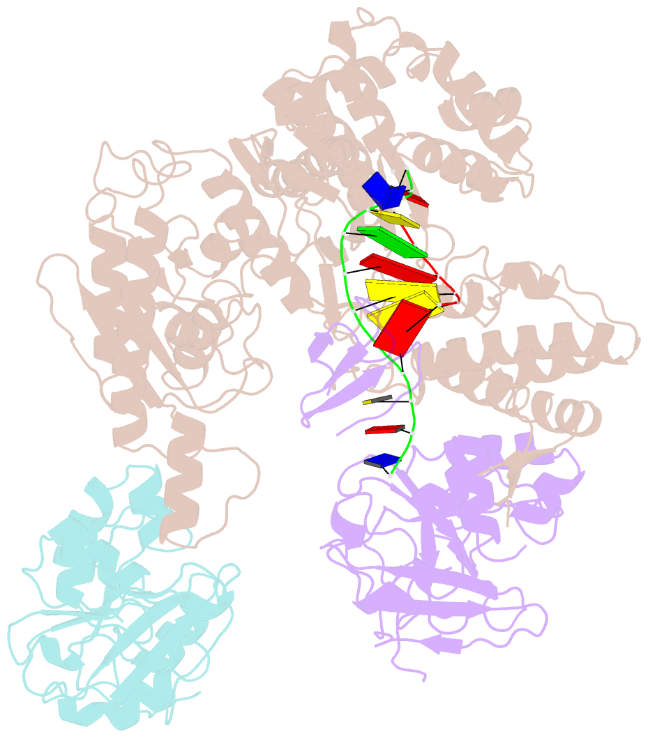
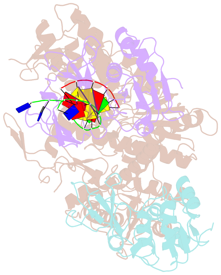
- Structure of bacteriophage t7 lagging-strand DNA polymerase (d5a-e7a) interacting with primase domains of two gp4 subunits bound to an RNA-DNA hybrid and dttp (from lags1)
- Reference
- Gao Y, Cui Y, Fox T, Lin S, Wang H, de Val N, Zhou ZH, Yang W (2019): "Structures and operating principles of the replisome." Science, 363. doi: 10.1126/science.aav7003.
- Abstract
- Visualization in atomic detail of the replisome that performs concerted leading- and lagging-DNA strand synthesis at a replication fork has not been reported. Using bacteriophage T7 as a model system, we determined cryo-electron microscopy structures up to 3.2-angstroms resolution of helicase translocating along DNA and of helicase-polymerase-primase complexes engaging in synthesis of both DNA strands. Each domain of the spiral-shaped hexameric helicase translocates sequentially hand-over-hand along a single-stranded DNA coil, akin to the way AAA+ ATPases (adenosine triphosphatases) unfold peptides. Two lagging-strand polymerases are attached to the primase, ready for Okazaki fragment synthesis in tandem. A β hairpin from the leading-strand polymerase separates two parental DNA strands into a T-shaped fork, thus enabling the closely coupled helicase to advance perpendicular to the downstream DNA duplex. These structures reveal the molecular organization and operating principles of a replisome.