Summary information and primary citation
- PDB-id
- 6uph; SNAP-derived features in text and JSON formats;
DNAproDB
- Class
- cell cycle
- Method
- cryo-EM (2.7 Å)
- Summary
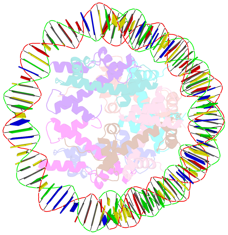
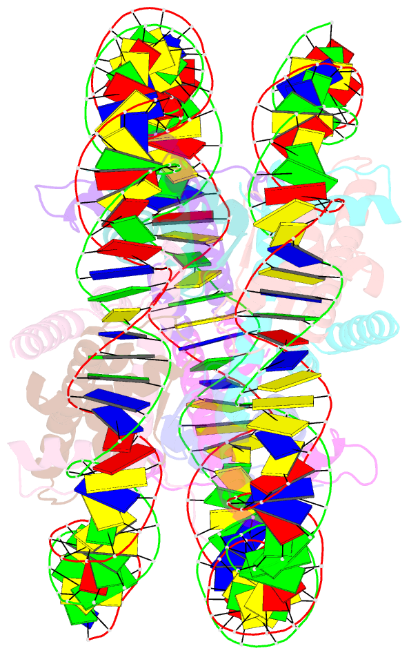
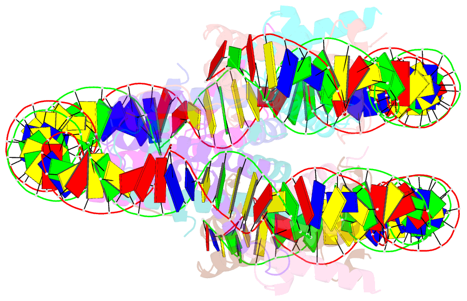
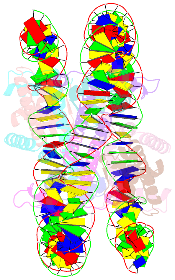
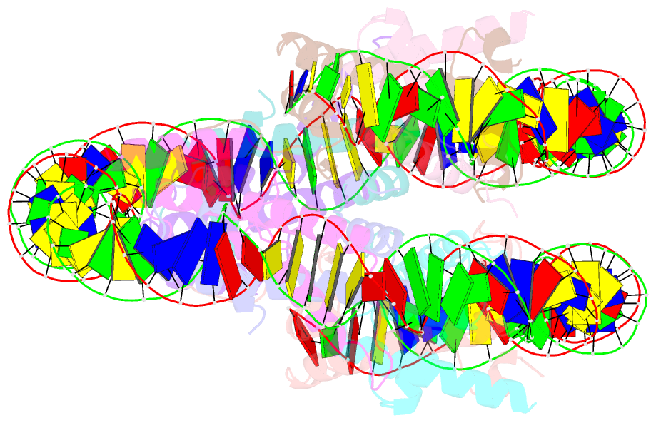
- Structure of a yeast centromeric nucleosome at 2.7 angstrom resolution
- Reference
- Migl D, Kschonsak M, Arthur CP, Khin Y, Harrison SC, Ciferri C, Dimitrova YN (2020): "Cryoelectron Microscopy Structure of a Yeast Centromeric Nucleosome at 2.7 angstrom Resolution." Structure, 28, 363-370.e3. doi: 10.1016/j.str.2019.12.002.
- Abstract
- Kinetochores mediate chromosome segregation during cell division. They assemble on centromeric nucleosomes and capture spindle microtubules. In budding yeast, a kinetochore links a single nucleosome, containing the histone variant Cse4CENP-A instead of H3, with a single microtubule. Conservation of most kinetochore components from yeast to metazoans suggests that the yeast kinetochore represents a module of the more complex metazoan arrangements. We describe here a streamlined protocol for reconstituting a yeast centromeric nucleosome and a systematic exploration of cryo-grid preparation. These developments allowed us to obtain a high-resolution cryoelectron microscopy reconstruction. As suggested by previous work, fewer base pairs are in tight association with the histone octamer than there are in canonical nucleosomes. Weak binding of the end DNA sequences may contribute to specific recognition by other inner kinetochore components. The centromeric nucleosome structure and the strategies we describe will facilitate studies of many other aspects of kinetochore assembly and chromatin biochemistry.