Summary information and primary citation
- PDB-id
- 7ub2; SNAP-derived features in text and JSON formats;
DNAproDB
- Class
- DNA binding protein-DNA
- Method
- cryo-EM (3.4 Å)
- Summary
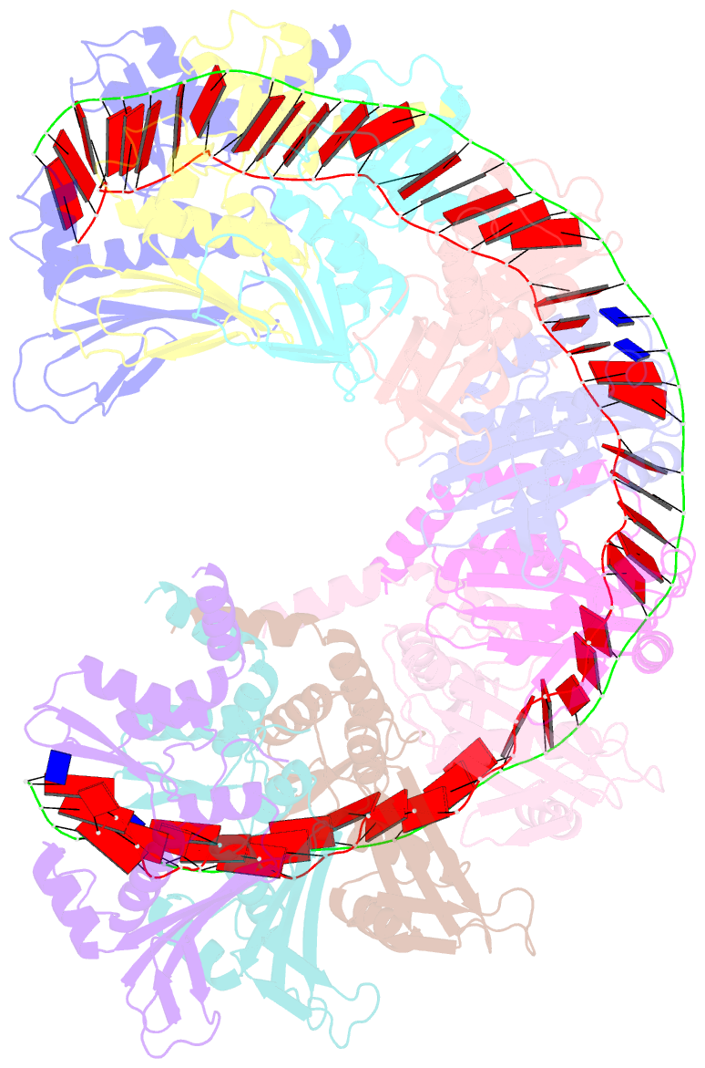
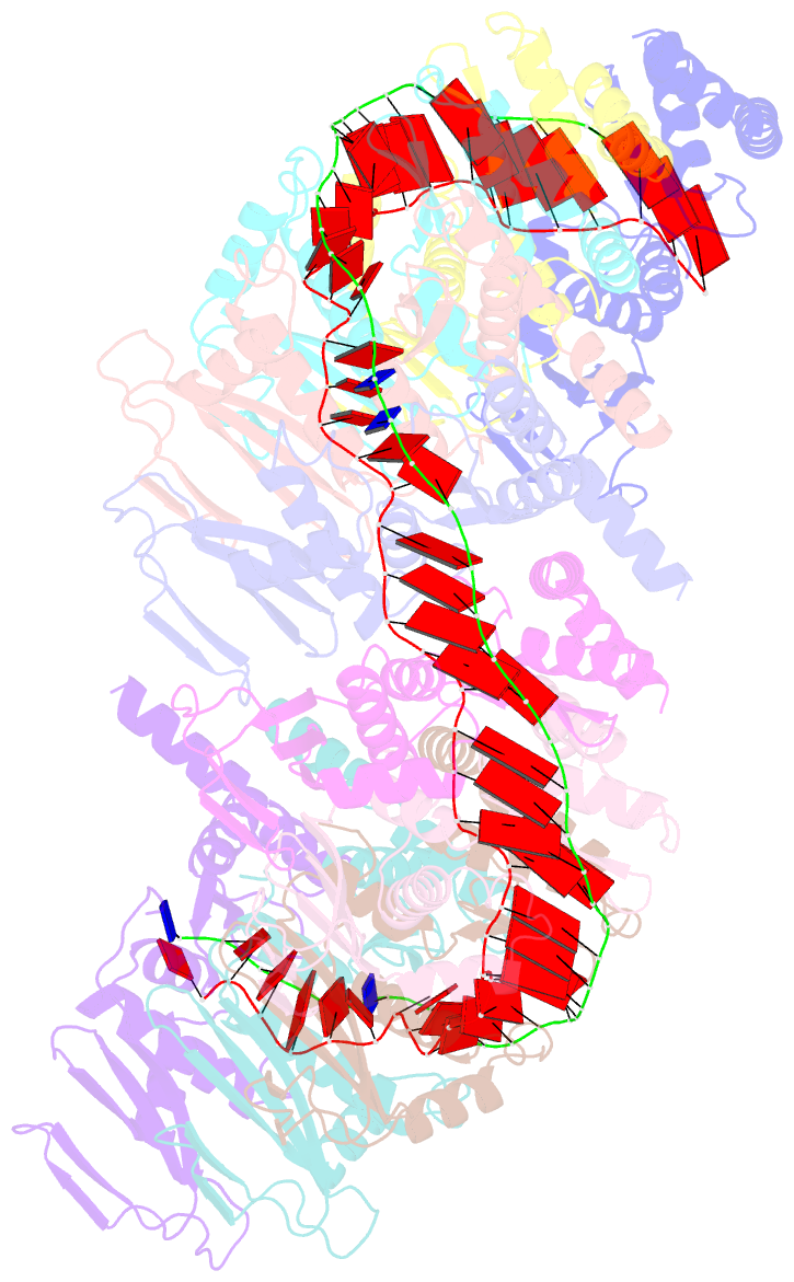
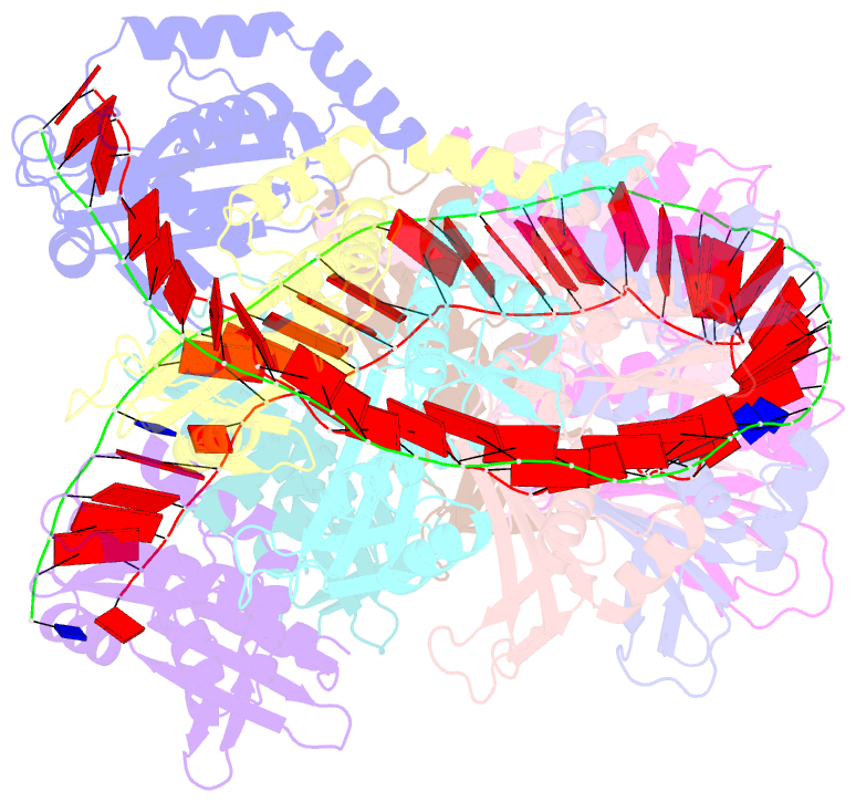
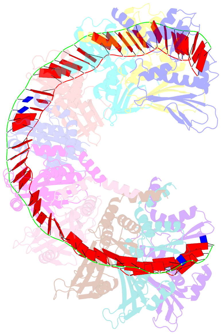
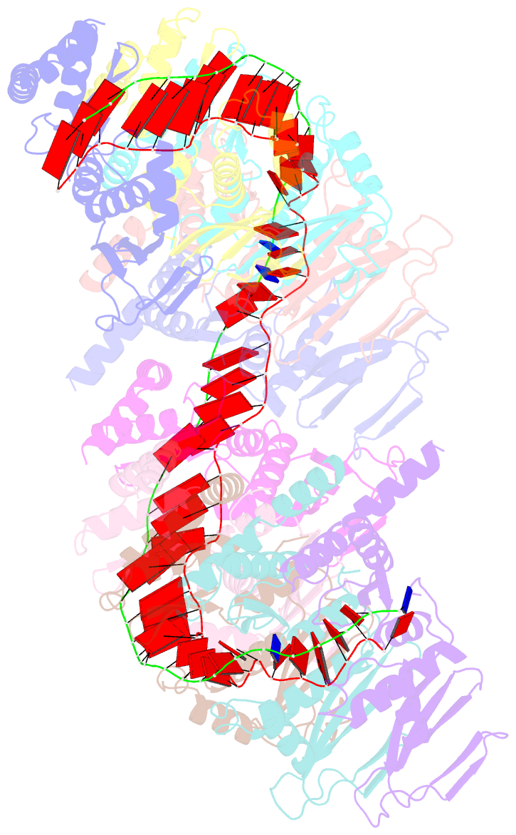
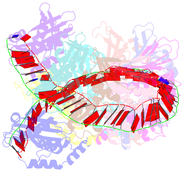
- Structure of rect protein from listeria innoccua phage a118 in complex with 83-mer annealed duplex
- Reference
- Caldwell BJ, Norris AS, Karbowski CF, Wiegand AM, Wysocki VH, Bell CE (2022): "Structure of a RecT/Red beta family recombinase in complex with a duplex intermediate of DNA annealing." Nat Commun, 13, 7855. doi: 10.1038/s41467-022-35572-z.
- Abstract
- Some bacteriophage encode a recombinase that catalyzes single-stranded DNA annealing (SSA). These proteins are apparently related to RAD52, the primary human SSA protein. The best studied protein, Redβ from bacteriophage λ, binds weakly to ssDNA, not at all to dsDNA, but tightly to a duplex intermediate of annealing formed when two complementary DNA strands are added to the protein sequentially. We used single particle cryo-electron microscopy (cryo-EM) to determine a 3.4 Å structure of a Redβ homolog from a prophage of Listeria innocua in complex with two complementary 83mer oligonucleotides. The structure reveals a helical protein filament bound to a DNA duplex that is highly extended and unwound. Native mass spectrometry confirms that the complex seen by cryo-EM is the predominant species in solution. The protein shares a common core fold with RAD52 and a similar mode of ssDNA-binding. These data provide insights into the mechanism of protein-catalyzed SSA.