Summary information and primary citation
- PDB-id
- 7ujl; SNAP-derived features in text and JSON formats;
DNAproDB
- Class
- recombination-DNA
- Method
- cryo-EM (3.3 Å)
- Summary
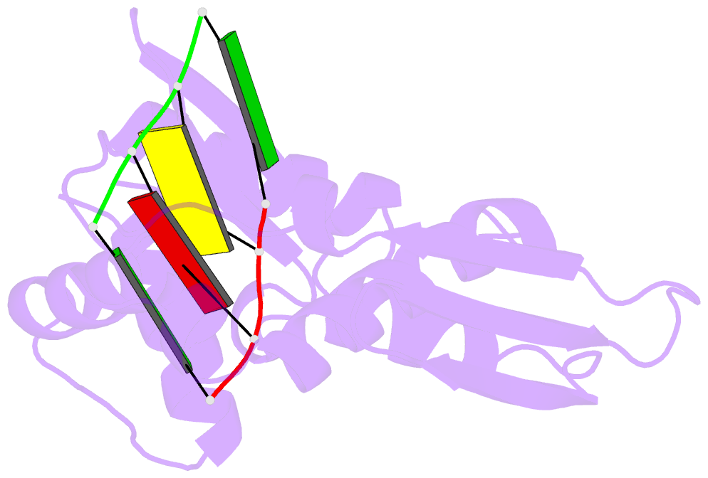
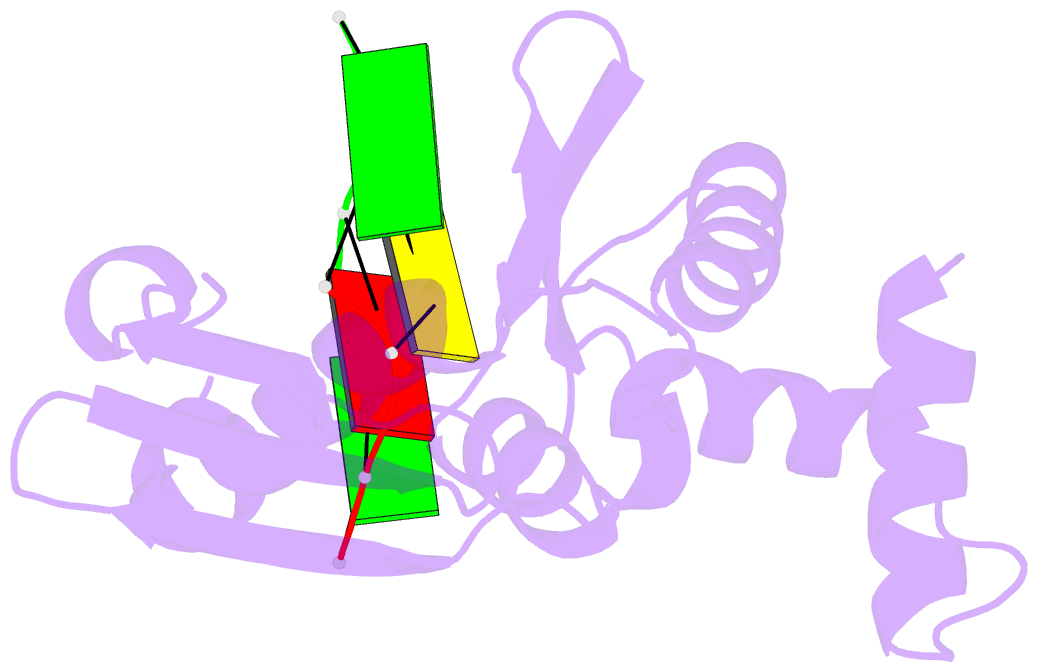
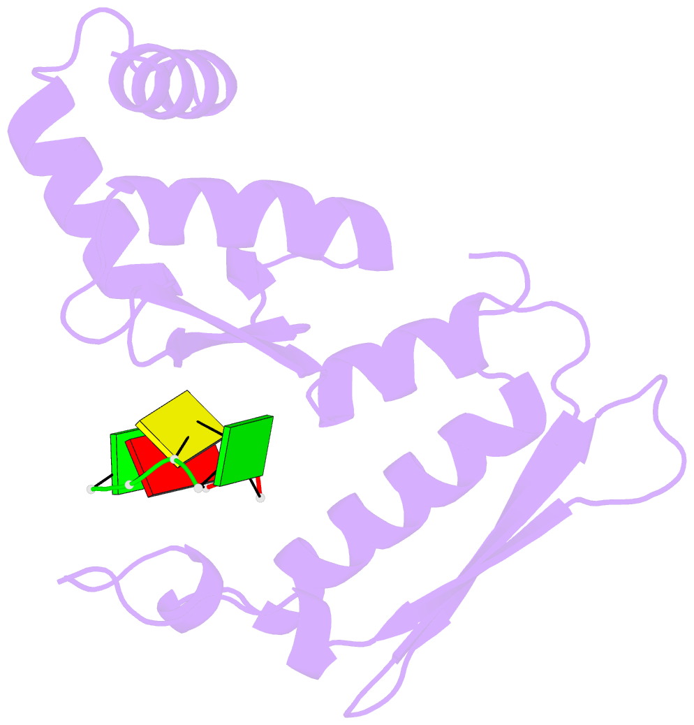
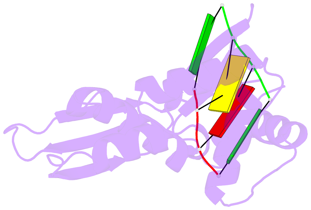
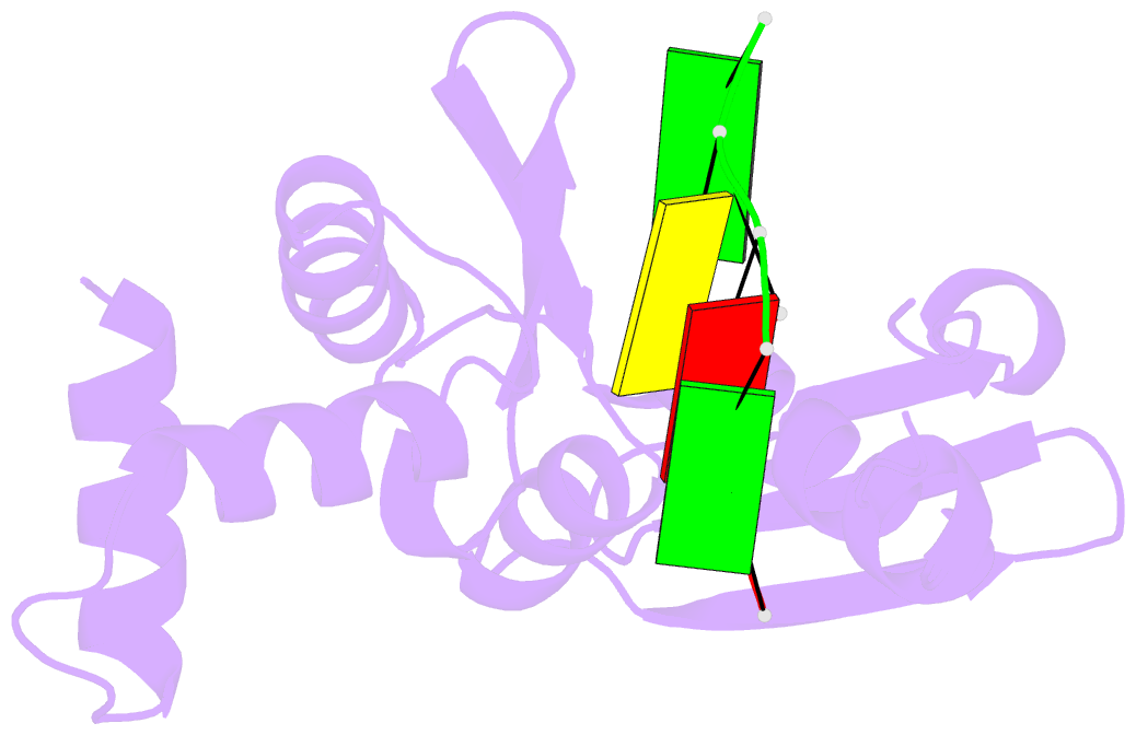
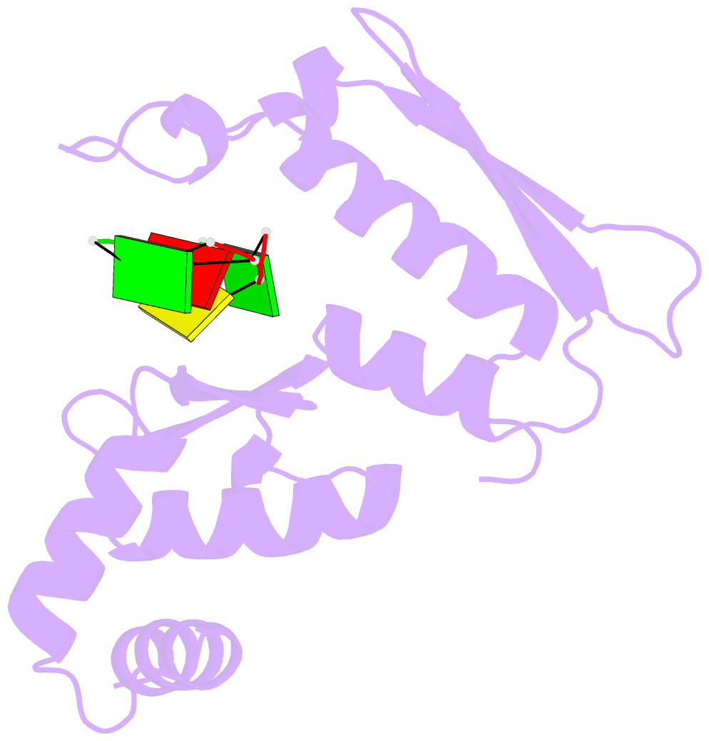
- Bacteriophage lambda red-beta n-terminal domain helical assembly in complex with dsDNA
- Reference
- Newing TP, Brewster JL, Fitschen LJ, Bouwer JC, Johnston NP, Yu H, Tolun G (2022): "Red beta 177 annealase structure reveals details of oligomerization and lambda Red-mediated homologous DNA recombination." Nat Commun, 13, 5649. doi: 10.1038/s41467-022-33090-6.
- Abstract
- The Redβ protein of the bacteriophage λ red recombination system is a model annealase which catalyzes single-strand annealing homologous DNA recombination. Here we present the structure of a helical oligomeric annealing intermediate of Redβ, consisting of N-terminal residues 1-177 bound to two complementary 27mer oligonucleotides, determined via cryogenic electron microscopy (cryo-EM) to a final resolution of 3.3 Å. The structure reveals a continuous binding groove which positions and stabilizes complementary DNA strands in a planar orientation to facilitate base pairing via a network of hydrogen bonding. Definition of the inter-subunit interface provides a structural basis for the propensity of Redβ to oligomerize into functionally significant long helical filaments, a trait shared by most annealases. Our cryo-EM structure and molecular dynamics simulations suggest that residues 133-138 form a flexible loop which modulates access to the binding groove. More than half a century after its discovery, this combination of structural and computational observations has allowed us to propose molecular mechanisms for the actions of the model annealase Redβ, a defining member of the Redβ/RecT protein family.