Summary information and primary citation
- PDB-id
- 8c7u; SNAP-derived features in text and JSON formats;
DNAproDB
- Class
- transcription
- Method
- X-ray (3.15 Å)
- Summary
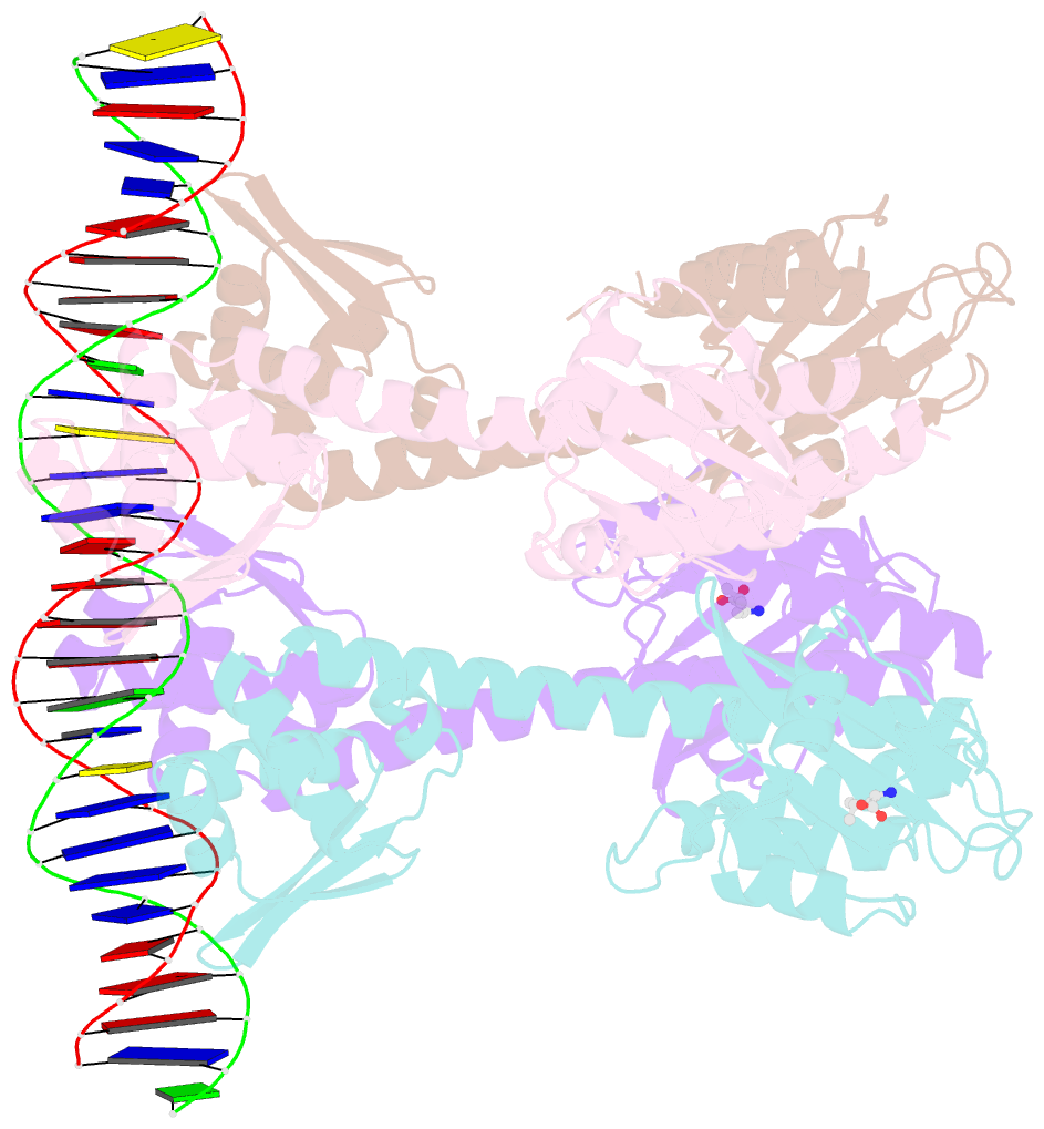
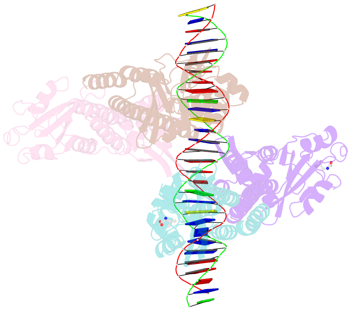
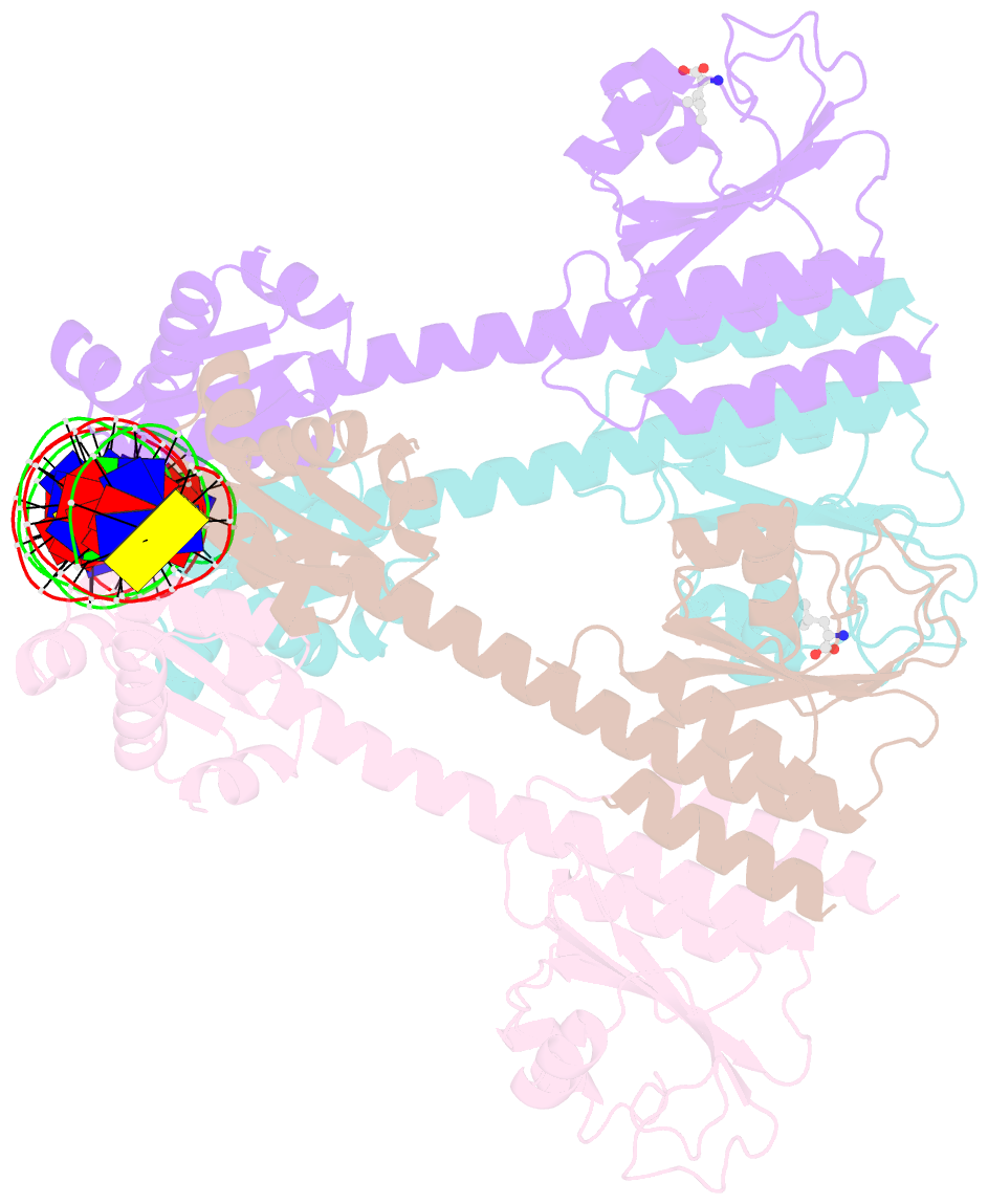
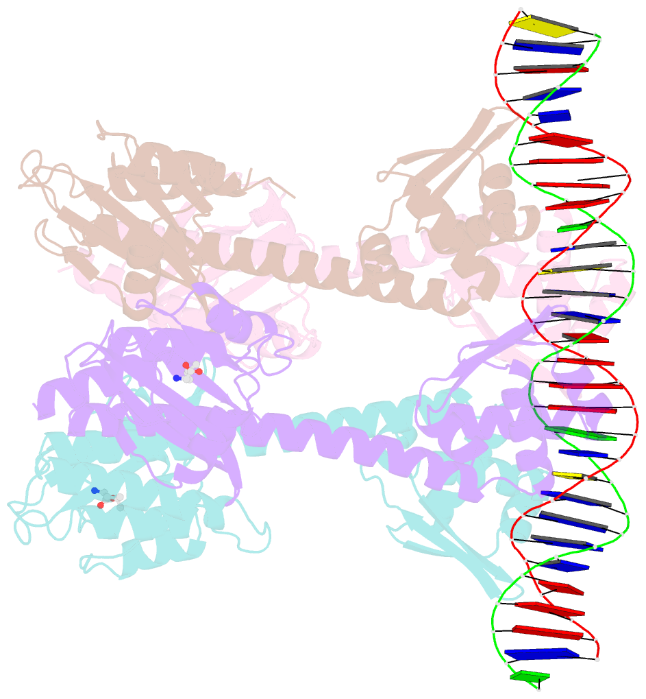
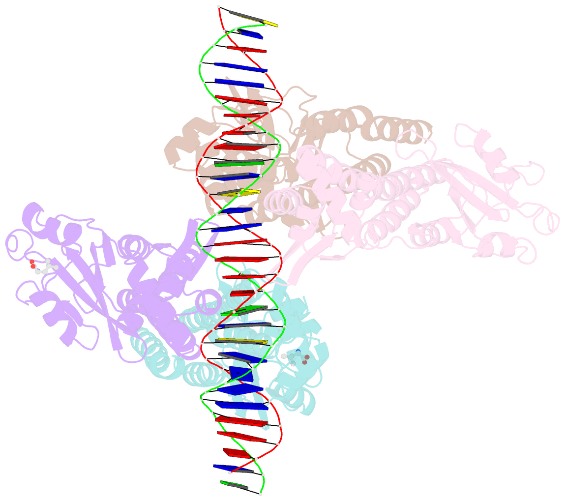
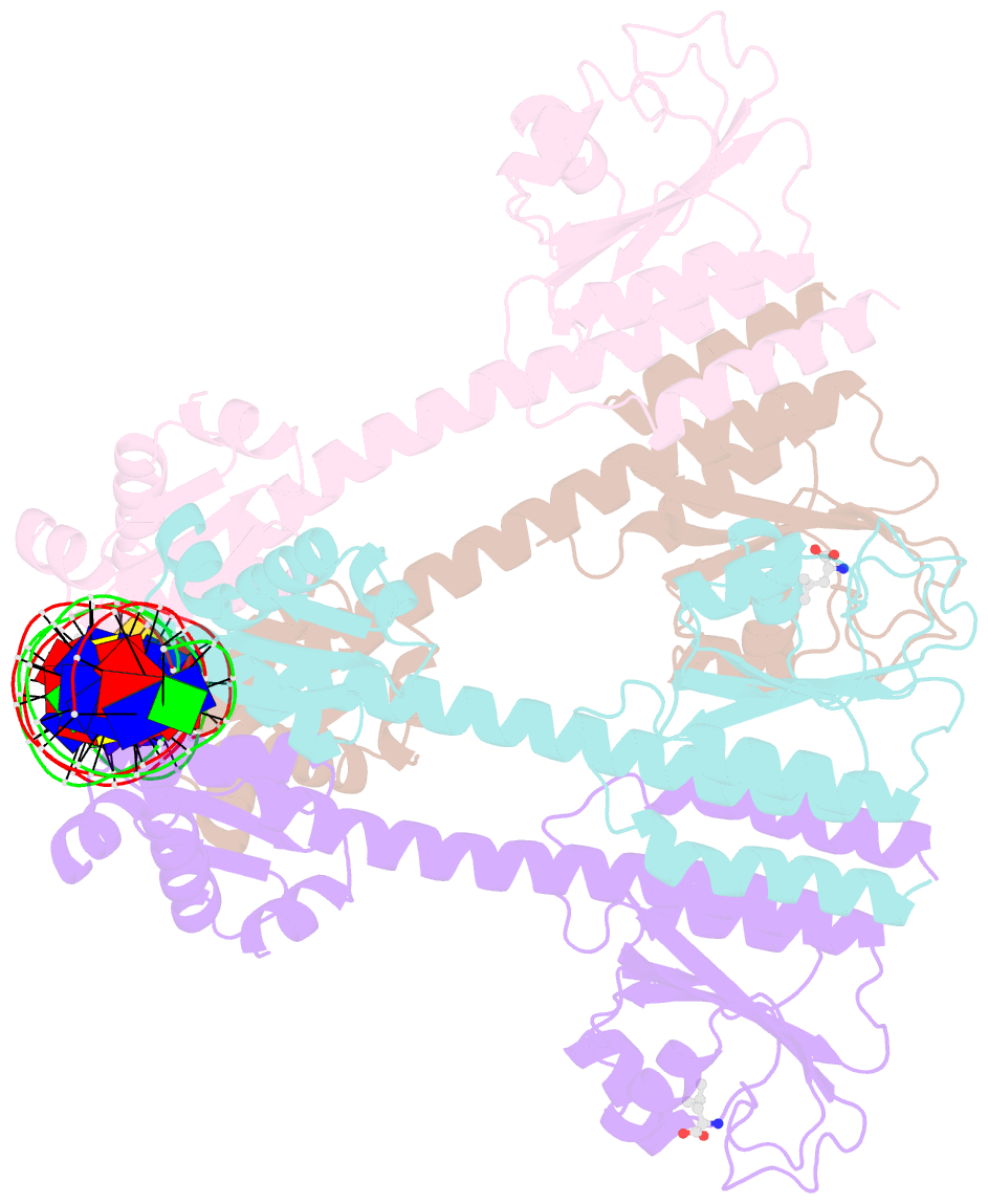
- Transcriptional pleiotropic repressor cody from enterococcus faecalis in complex with leu and a 30-bp DNA fragment encompassing two overlapping binding sites
- Reference
- Hainzl T, Bonde M, Almqvist F, Johansson J, Sauer-Eriksson AE (2023): "Structural insights into CodY activation and DNA recognition." Nucleic Acids Res., 51, 7631-7648. doi: 10.1093/nar/gkad512.
- Abstract
- Virulence factors enable pathogenic bacteria to infect host cells, establish infection, and contribute to disease progressions. In Gram-positive pathogens such as Staphylococcus aureus (Sa) and Enterococcus faecalis (Ef), the pleiotropic transcription factor CodY plays a key role in integrating metabolism and virulence factor expression. However, to date, the structural mechanisms of CodY activation and DNA recognition are not understood. Here, we report the crystal structures of CodY from Sa and Ef in their ligand-free form and their ligand-bound form complexed with DNA. Binding of the ligands-branched chain amino acids and GTP-induces conformational changes in the form of helical shifts that propagate to the homodimer interface and reorient the linker helices and DNA binding domains. DNA binding is mediated by a non-canonical recognition mechanism dictated by DNA shape readout. Furthermore, two CodY dimers bind to two overlapping binding sites in a highly cooperative manner facilitated by cross-dimer interactions and minor groove deformation. Our structural and biochemical data explain how CodY can bind a wide range of substrates, a hallmark of many pleiotropic transcription factors. These data contribute to a better understanding of the mechanisms underlying virulence activation in important human pathogens.