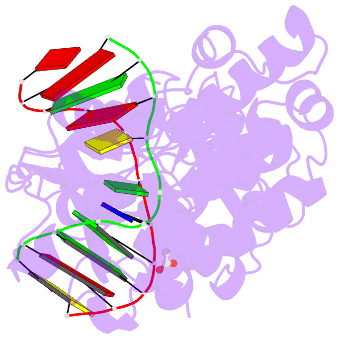
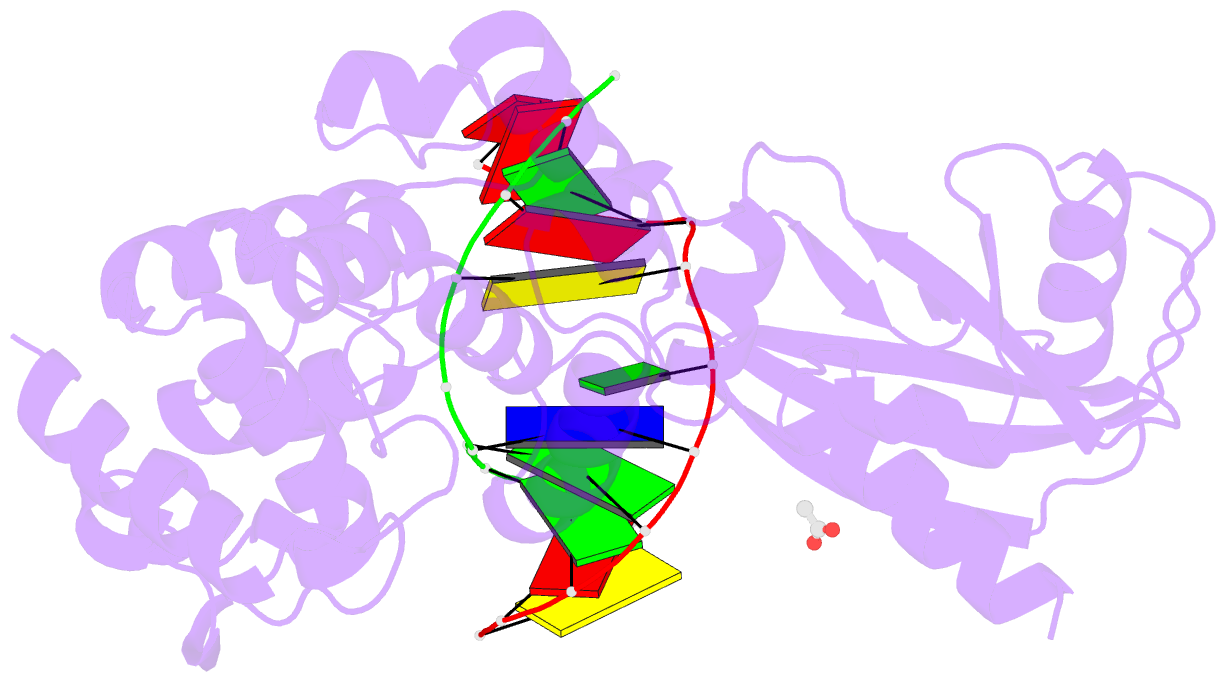
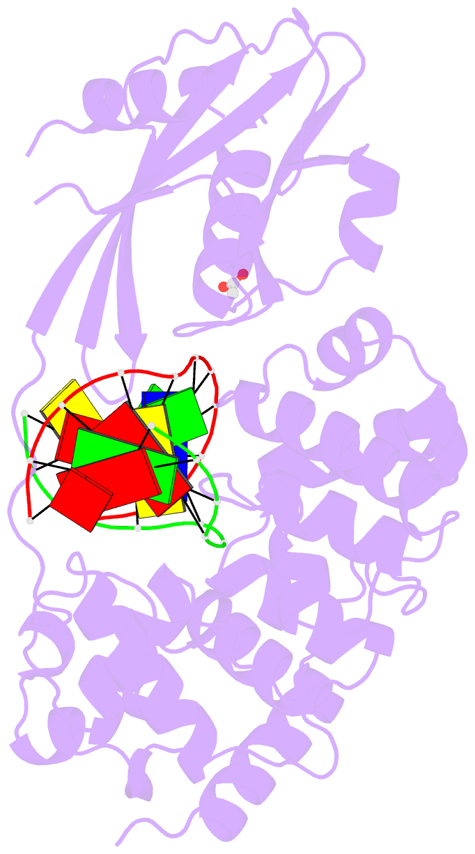
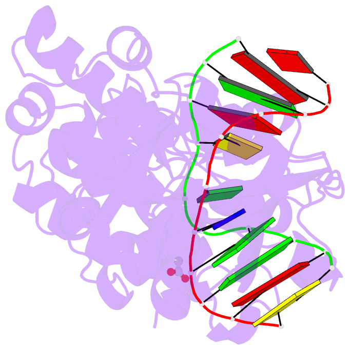
Summary information and primary citation
- PDB-id
- 8dvy; SNAP-derived features in text and JSON formats;
DNAproDB
- Class
- hydrolase-DNA
- Method
- X-ray (2.36 Å)
- Summary
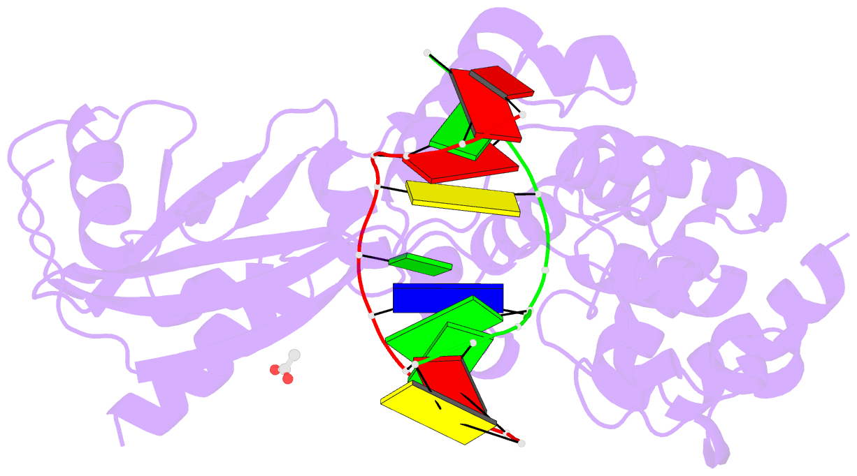
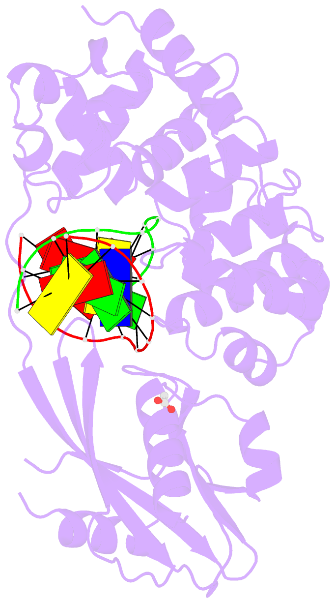
- DNA glycosylase muty variant n146s in complex with DNA containing d(8-oxo-g) paired with an enzyme-generated abasic site product (ap) and crystalized with calcium acetate
- Reference
- Demir M, Russelburg LP, Lin WJ, Trasvina-Arenas CH, Huang B, Yuen PK, Horvath MP, David SS (2023): "Structural snapshots of base excision by the cancer-associated variant MutY N146S reveal a retaining mechanism." Nucleic Acids Res., 51, 1034-1049. doi: 10.1093/nar/gkac1246.
- Abstract
- DNA glycosylase MutY plays a critical role in suppression of mutations resulted from oxidative damage, as highlighted by cancer-association of the human enzyme. MutY requires a highly conserved catalytic Asp residue for excision of adenines misinserted opposite 8-oxo-7,8-dihydroguanine (OG). A nearby Asn residue hydrogen bonds to the catalytic Asp in structures of MutY and its mutation to Ser is an inherited variant in human MUTYH associated with colorectal cancer. We captured structural snapshots of N146S Geobacillus stearothermophilus MutY bound to DNA containing a substrate, a transition state analog and enzyme-catalyzed abasic site products to provide insight into the base excision mechanism of MutY and the role of Asn. Surprisingly, despite the ability of N146S to excise adenine and purine (P) in vitro, albeit at slow rates, N146S-OG:P complex showed a calcium coordinated to the purine base altering its conformation to inhibit hydrolysis. We obtained crystal structures of N146S Gs MutY bound to its abasic site product by removing the calcium from crystals of N146S-OG:P complex to initiate catalysis in crystallo or by crystallization in the absence of calcium. The product structures of N146S feature enzyme-generated β-anomer abasic sites that support a retaining mechanism for MutY-catalyzed base excision.