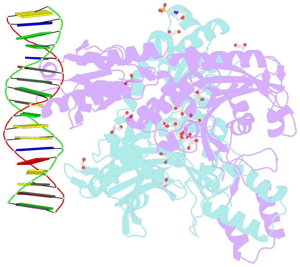
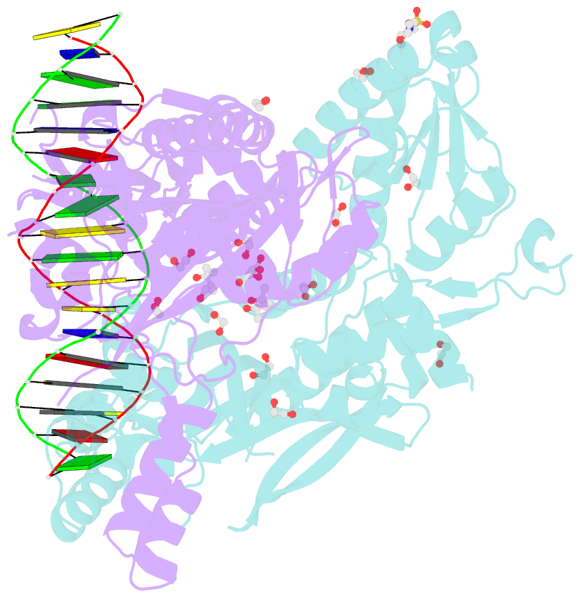
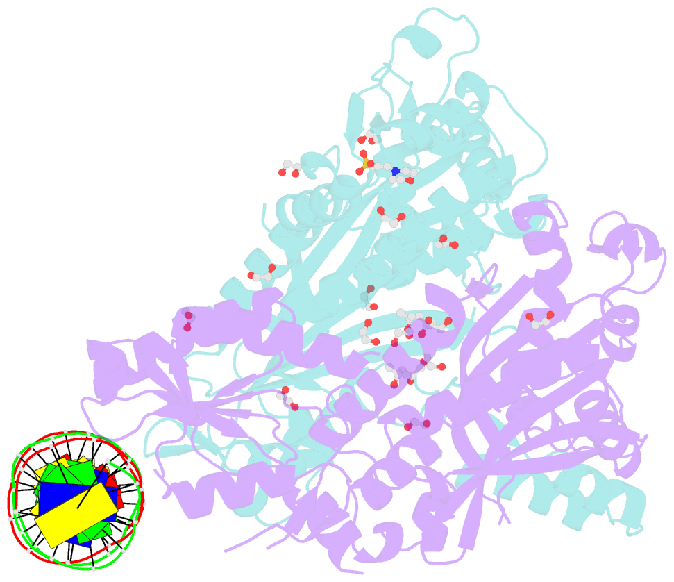
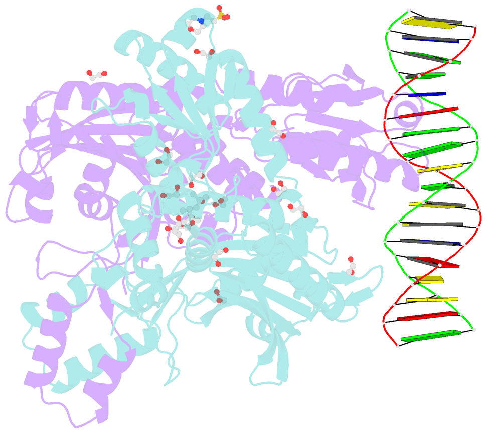
Summary information and primary citation
- PDB-id
- 8f69; SNAP-derived features in text and JSON formats;
DNAproDB
- Class
- DNA binding protein-DNA
- Method
- X-ray (2.2 Å)
- Summary
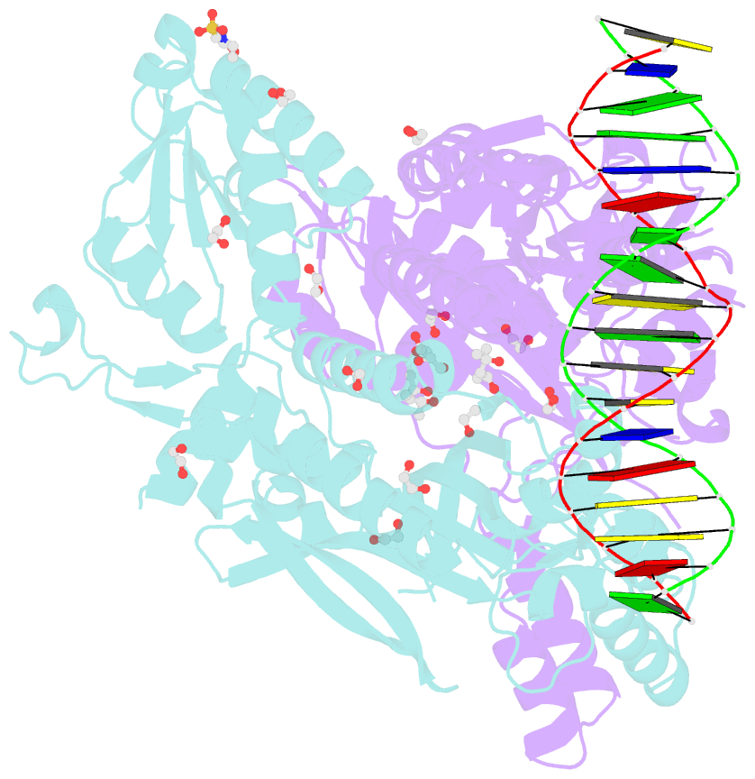
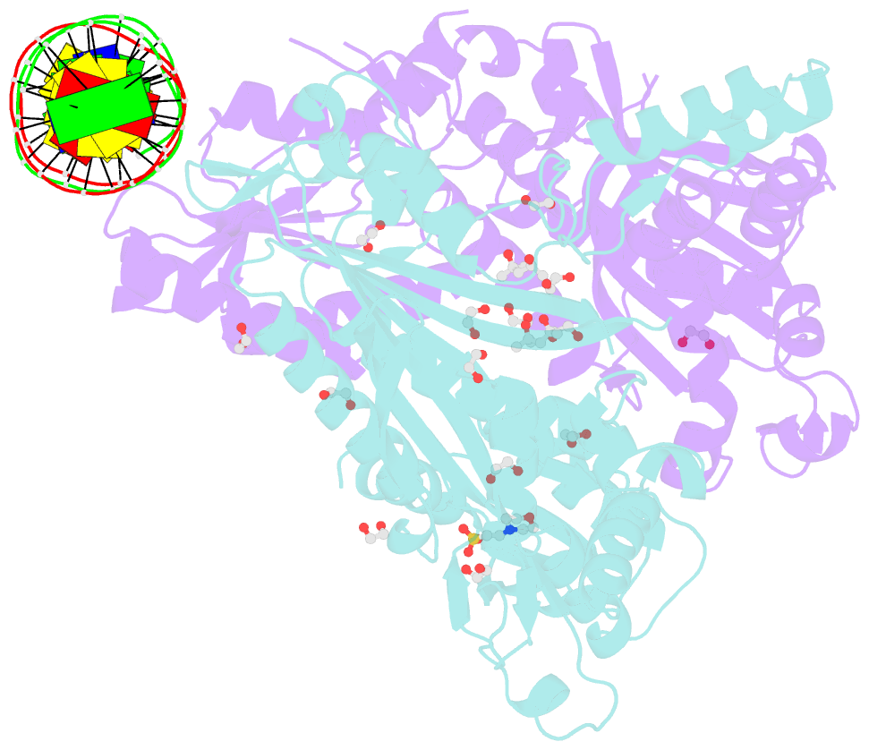
- Crystal structure of murine polg2 dimer bound to DNA
- Reference
- Wojtaszek JL, Hoff KE, Longley MJ, Kaur P, Andres SN, Wang H, Williams RS, Copeland WC (2023): "Structure-specific roles for PolG2-DNA complexes in maintenance and replication of mitochondrial DNA." Nucleic Acids Res., 51, 9716-9732. doi: 10.1093/nar/gkad679.
- Abstract
- The homodimeric PolG2 accessory subunit of the mitochondrial DNA polymerase gamma (Pol γ) enhances DNA binding and processive DNA synthesis by the PolG catalytic subunit. PolG2 also directly binds DNA, although the underlying molecular basis and functional significance are unknown. Here, data from Atomic Force Microscopy (AFM) and X-ray structures of PolG2-DNA complexes define dimeric and hexameric PolG2 DNA binding modes. Targeted disruption of PolG2 DNA-binding interfaces impairs processive DNA synthesis without diminishing Pol γ subunit affinities. In addition, a structure-specific DNA-binding role for PolG2 oligomers is supported by X-ray structures and AFM showing that oligomeric PolG2 localizes to DNA crossings and targets forked DNA structures resembling the mitochondrial D-loop. Overall, data indicate that PolG2 DNA binding has both PolG-dependent and -independent functions in mitochondrial DNA replication and maintenance, which provide new insight into molecular defects associated with PolG2 disruption in mitochondrial disease.