Summary information and primary citation
- PDB-id
- 8usn; SNAP-derived features in text and JSON formats;
DNAproDB
- Class
- viral protein
- Method
- cryo-EM (8.9 Å)
- Summary
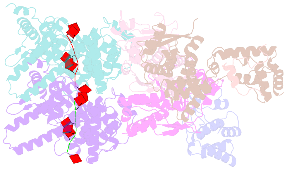
- Intracellular cryo-tomography structure of ebov nucleocapsid at 8.9 angstrom
- Reference
- Watanabe R, Zyla D, Parekh D, Hong C, Jones Y, Schendel SL, Wan W, Castillon G, Saphire EO (2024): "Intracellular Ebola virus nucleocapsid assembly revealed by in situ cryo-electron tomography." Cell, 187, 5587-5603.e19. doi: 10.1016/j.cell.2024.08.044.
- Abstract
- Filoviruses, including the Ebola and Marburg viruses, cause hemorrhagic fevers with up to 90% lethality. The viral nucleocapsid is assembled by polymerization of the nucleoprotein (NP) along the viral genome, together with the viral proteins VP24 and VP35. We employed cryo-electron tomography of cells transfected with viral proteins and infected with model Ebola virus to illuminate assembly intermediates, as well as a 9 Å map of the complete intracellular assembly. This structure reveals a previously unresolved third and outer layer of NP complexed with VP35. The intrinsically disordered region, together with the C-terminal domain of this outer layer of NP, provides the constant width between intracellular nucleocapsid bundles and likely functions as a flexible tether to the viral matrix protein in the virion. A comparison of intracellular nucleocapsids with prior in-virion nucleocapsid structures reveals that the nucleocapsid further condenses vertically in the virion. The interfaces responsible for nucleocapsid assembly are highly conserved and offer targets for broadly effective antivirals.


