Summary information and primary citation
- PDB-id
-
8ves;
DSSR-derived features in text and
JSON formats; DNAproDB
- Class
- hydrolase
- Method
- cryo-EM (3.22 Å)
- Summary
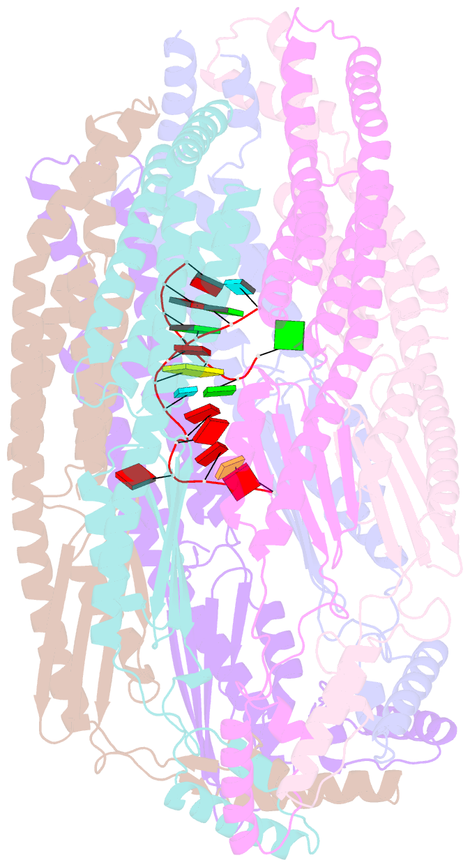
- Structure of yicc endoribonuclease bound to an RNA
substrate
- Reference
-
Wu R, Ingle S, Barnes SA, Dahlin HR, Khamrui S, Xiang Y,
Shi Y, Bechhofer DH, Lazarus MB (2024): "Structural
insights into RNA cleavage by a novel family of bacterial
RNases." Nucleic Acids Res.,
52, 10705-10716. doi: 10.1093/nar/gkae717.
- Abstract
- Processing of RNA is a key regulatory mechanism for all
living systems. Escherichia coli protein YicC belongs to
the well-conserved YicC family and has been identified as a
novel ribonuclease. Here, we report a 2.8-Å-resolution
crystal structure of the E. coli YicC apo protein and a
3.2-Å-cryo-EM structure of YicC bound to an RNA substrate.
The apo YicC forms a dimer of trimers with a large open
channel. In the RNA-bound form, the top trimer of YicC
rotates nearly 70° and closes the RNA substrate inside
the cavity to form a clamshell-pearl conformation that
resembles no other known RNases. The structural information
combined with mass spectrometry and biochemical data
identified cleavage on the upstream side of an RNA hairpin.
Mutagenesis studies demonstrated that the previously
uncharacterized domain, DUF1732, is critical in both RNA
binding and catalysis. These studies shed light on the
mechanism of the previously unexplored YicC RNase
family.


